Bulletin Board of Oral Pathology
Bulletin Board of Oral Pathology
|
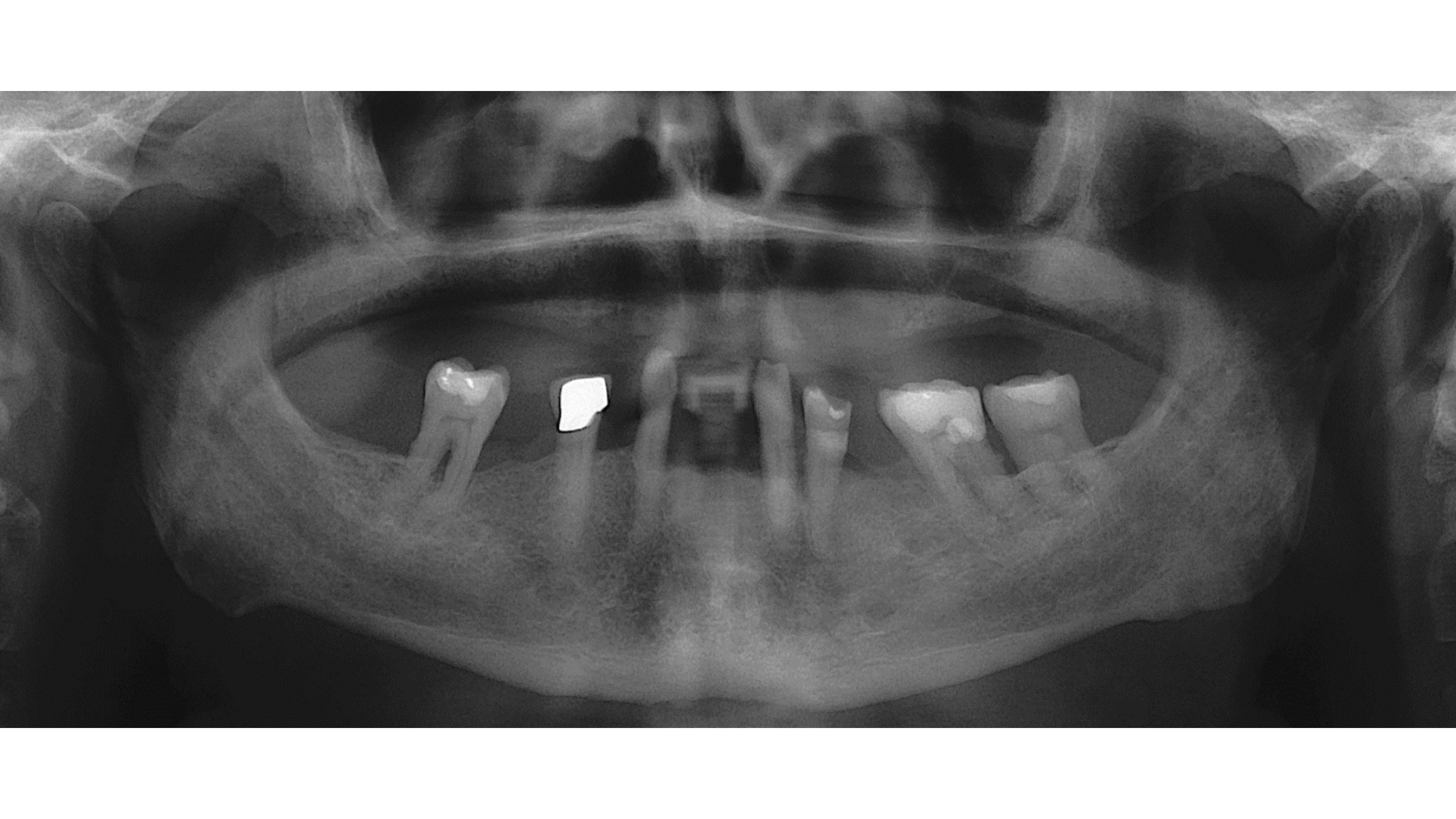
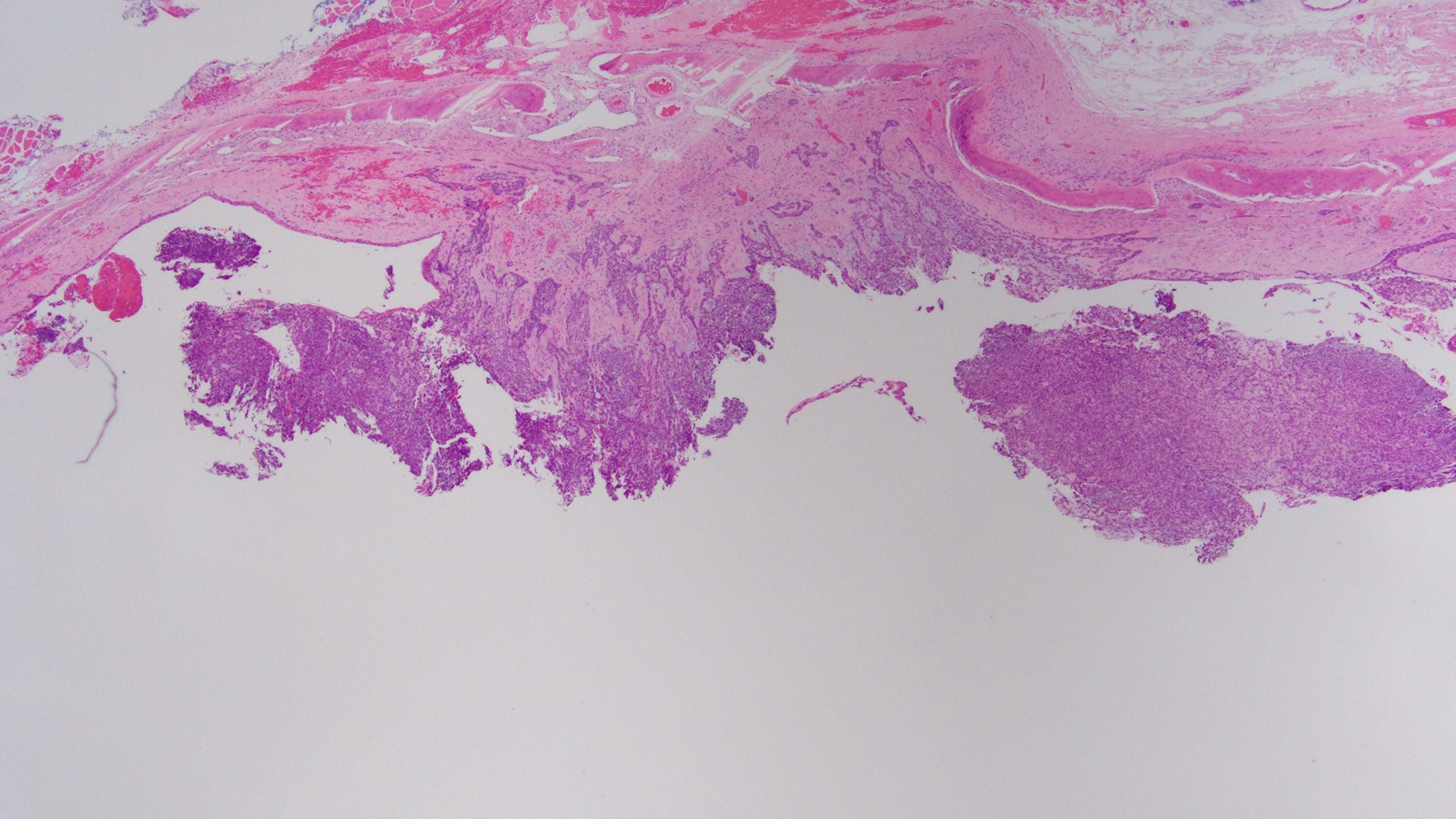
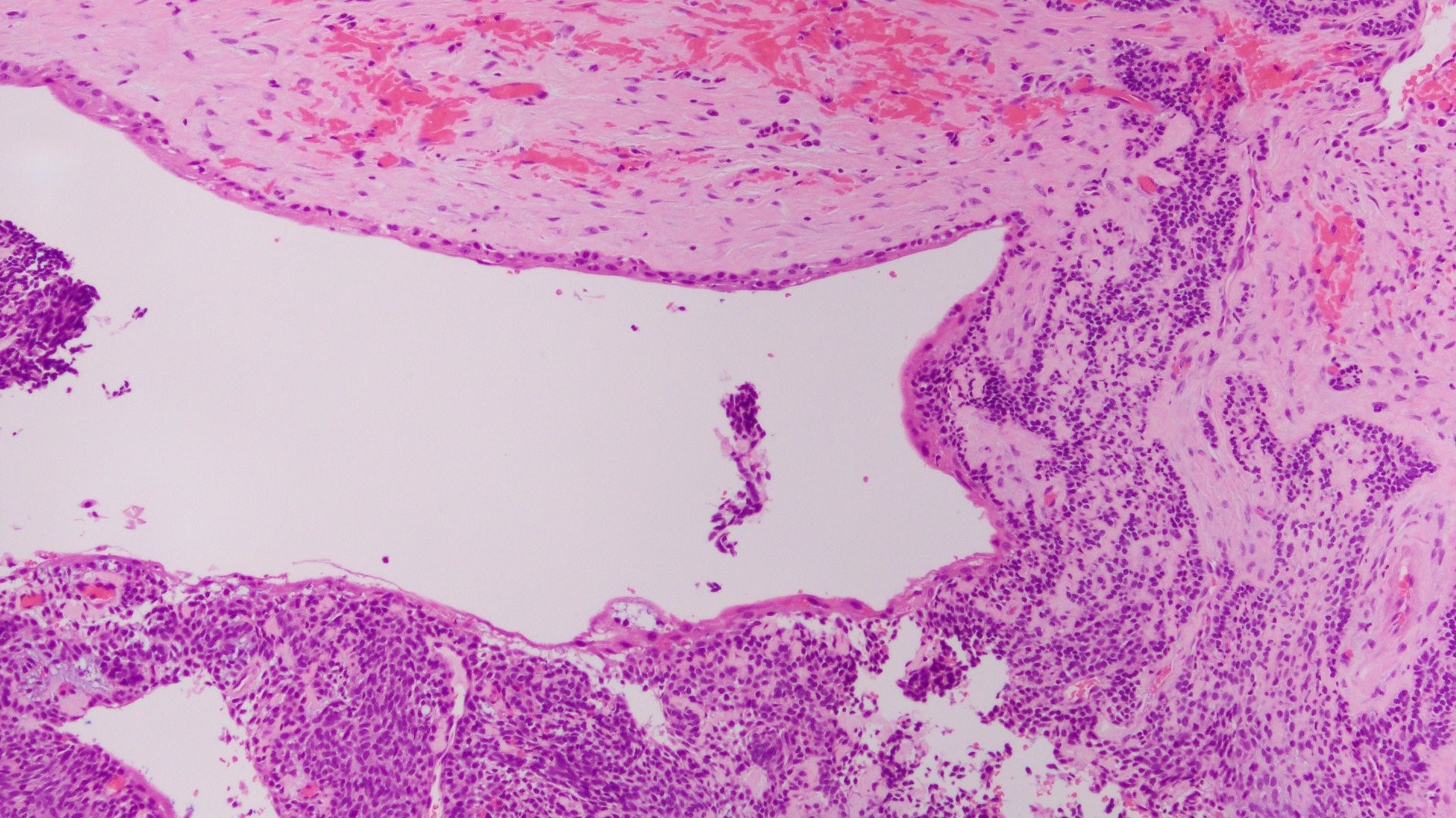
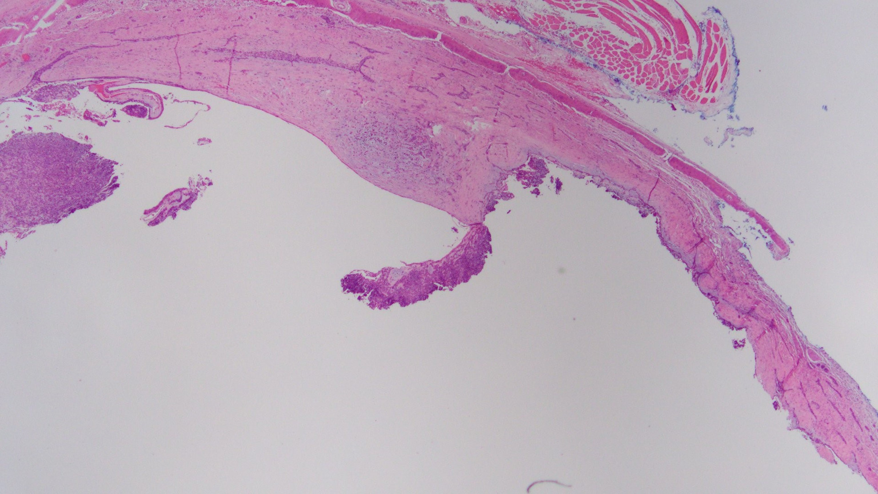
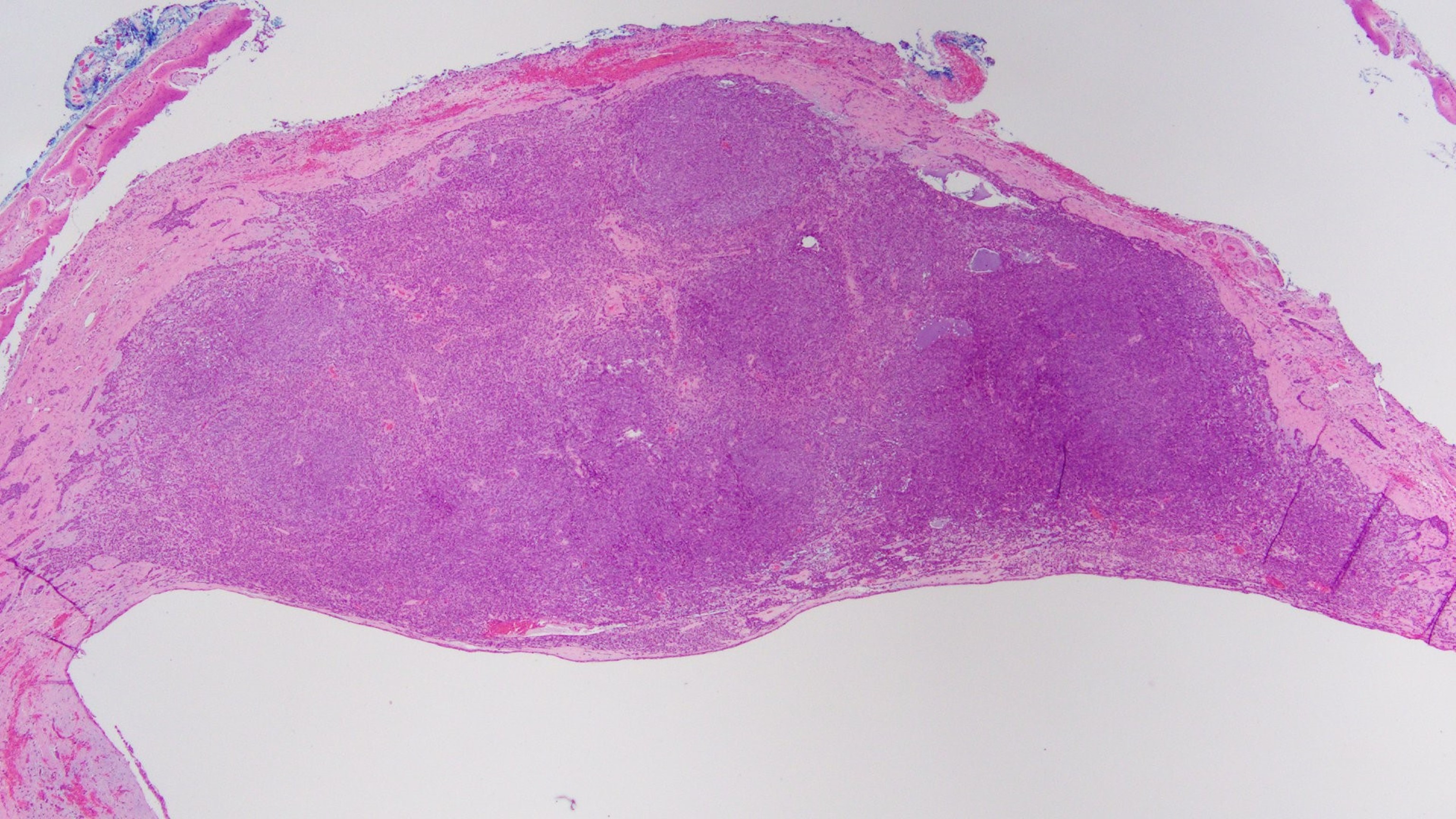
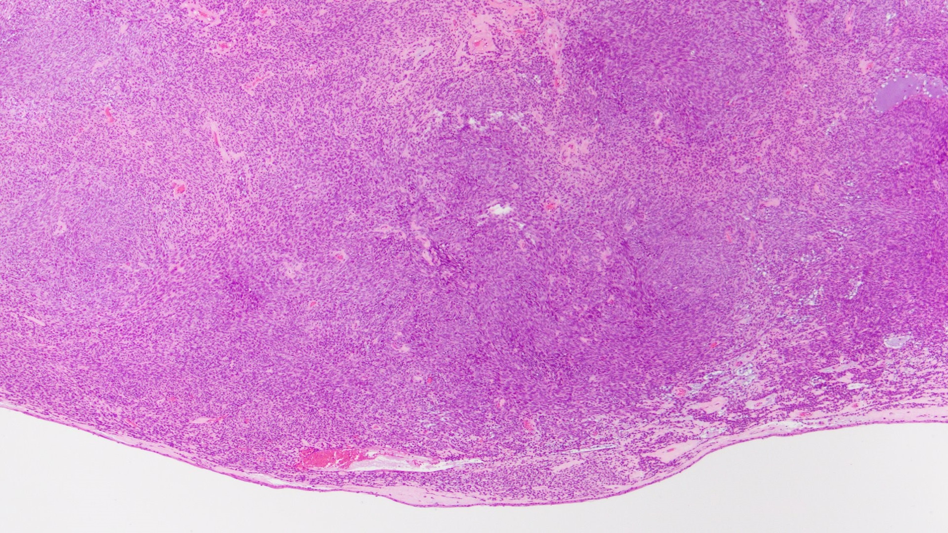
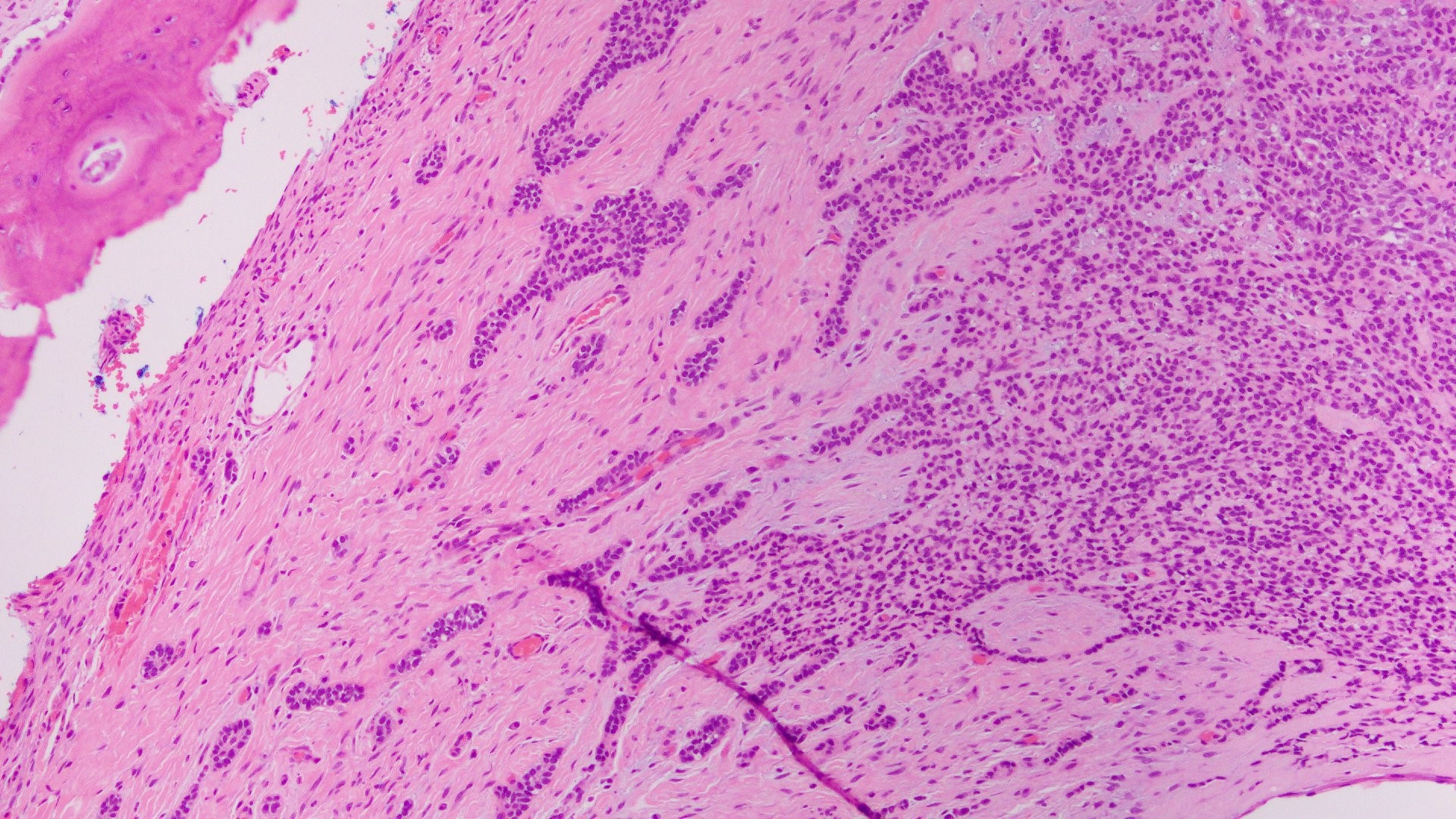
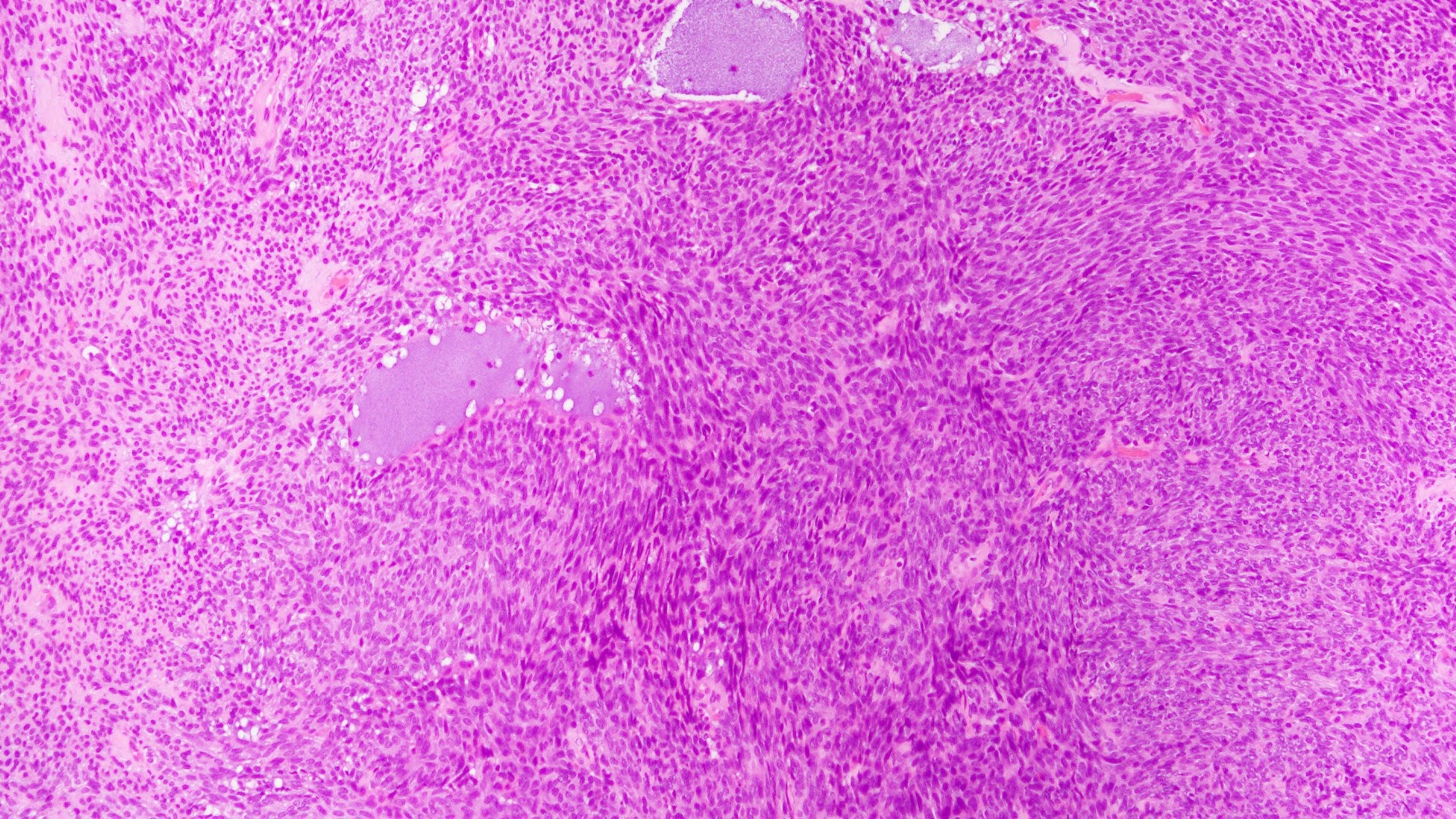
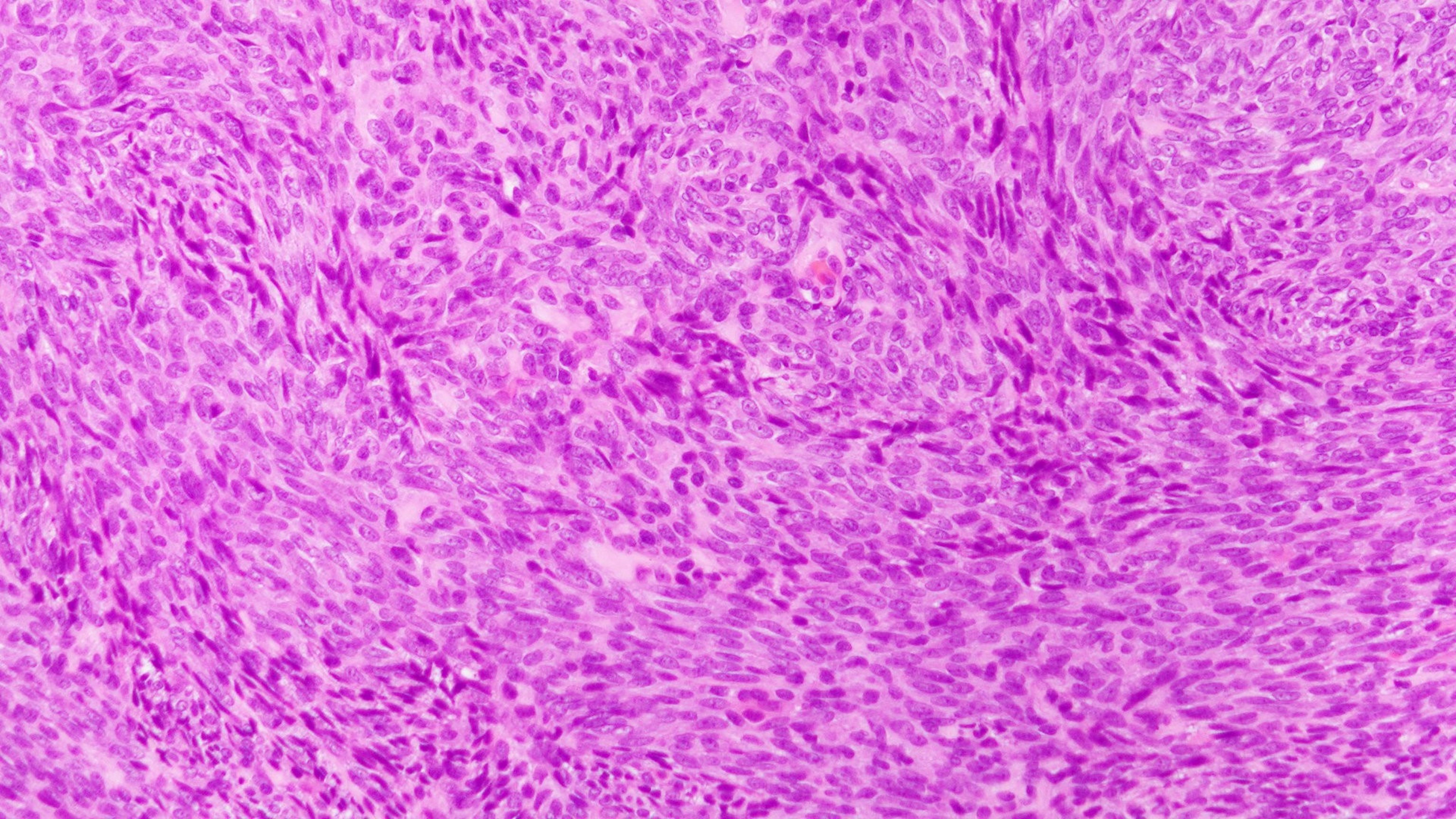
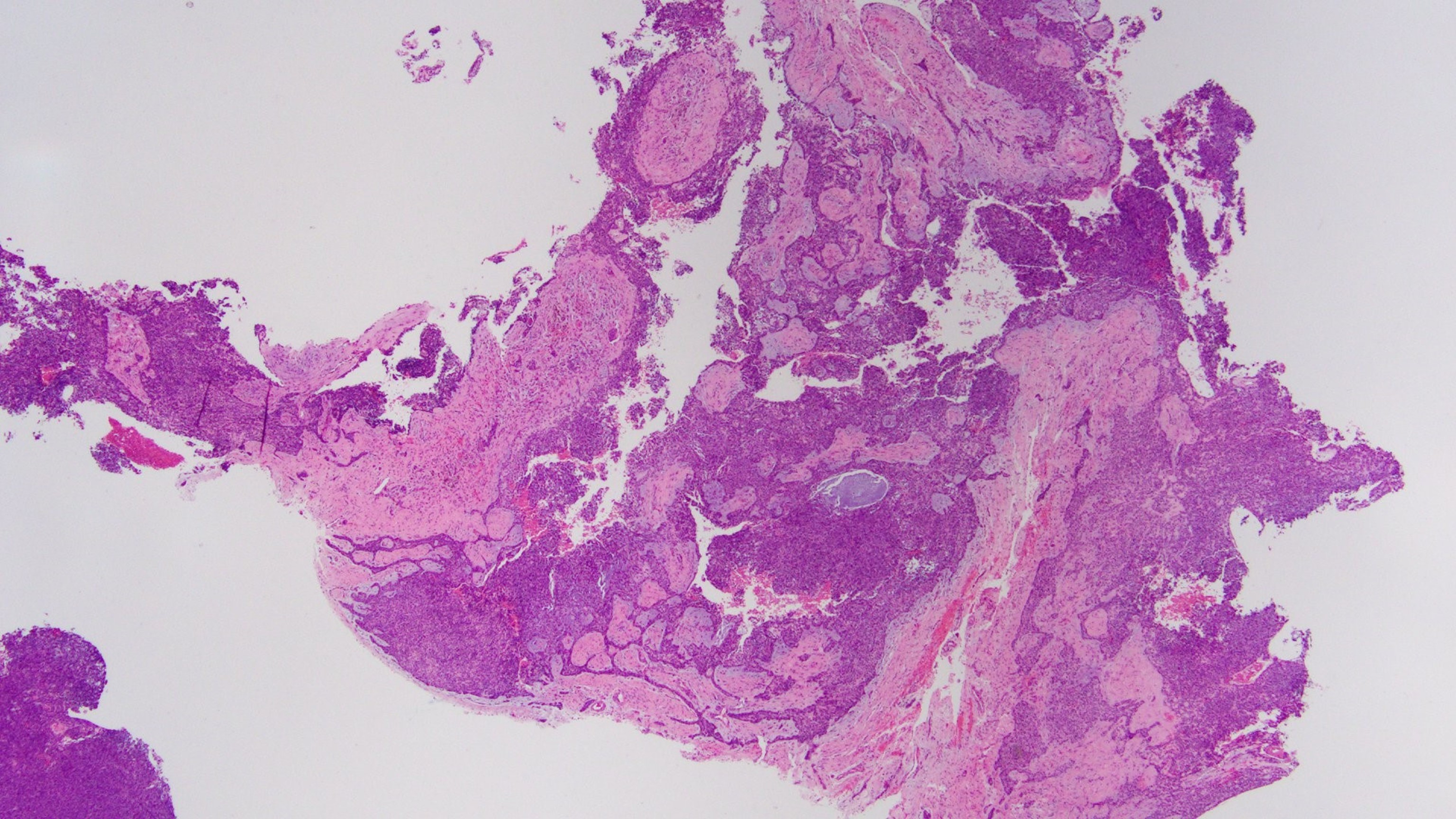
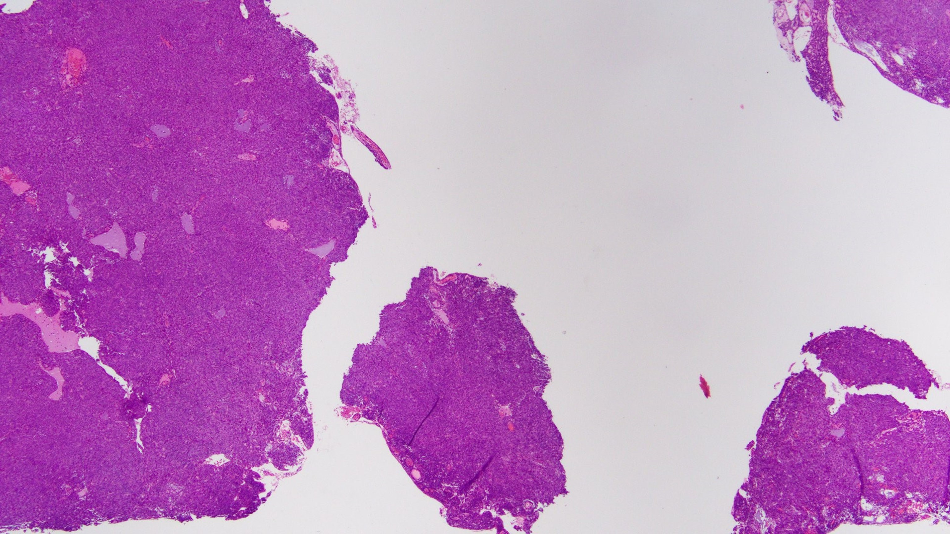
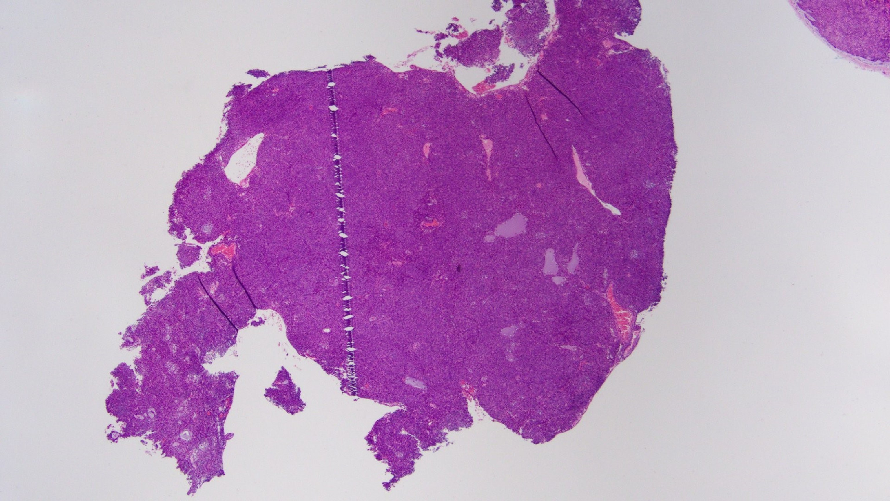
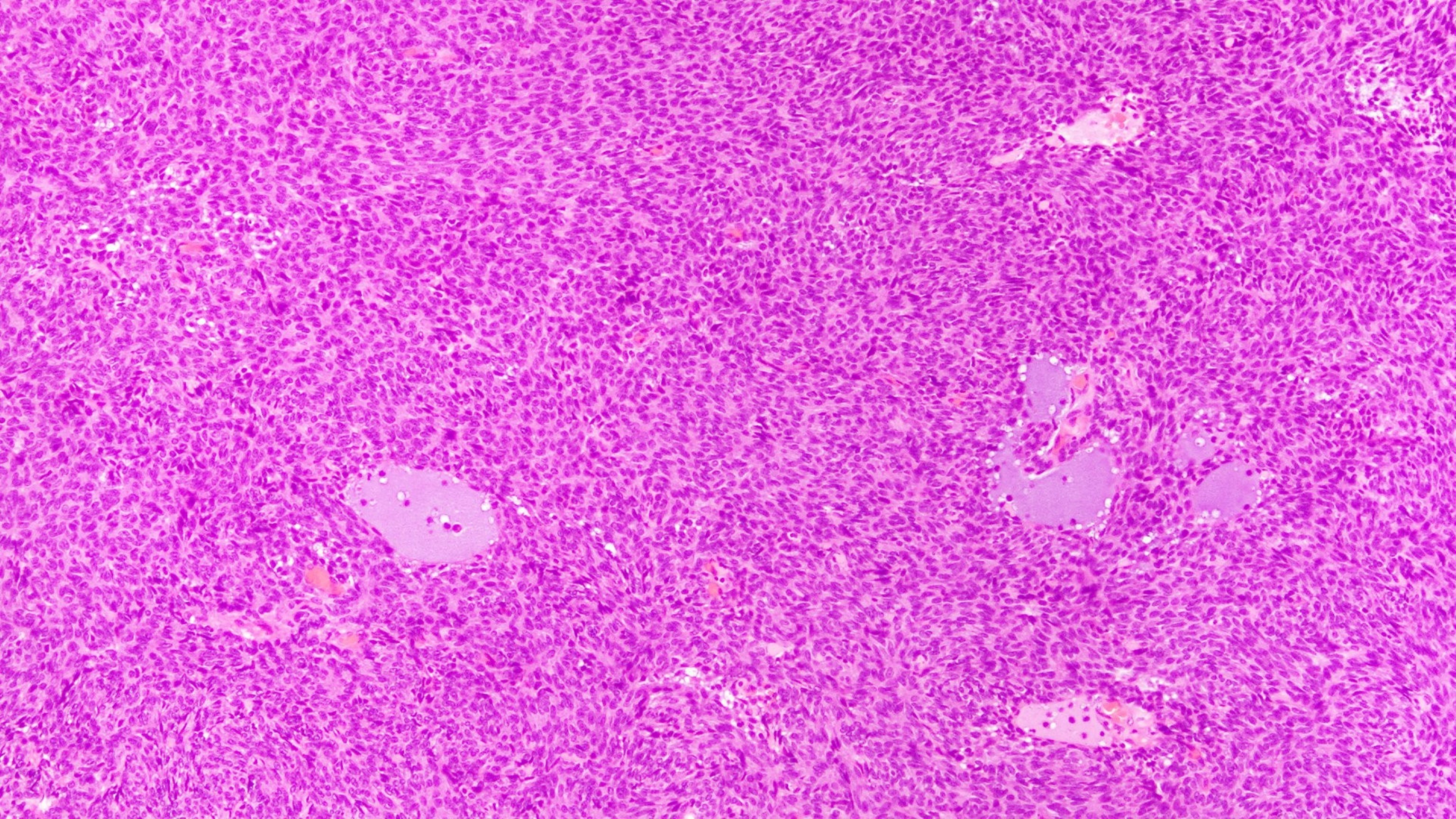
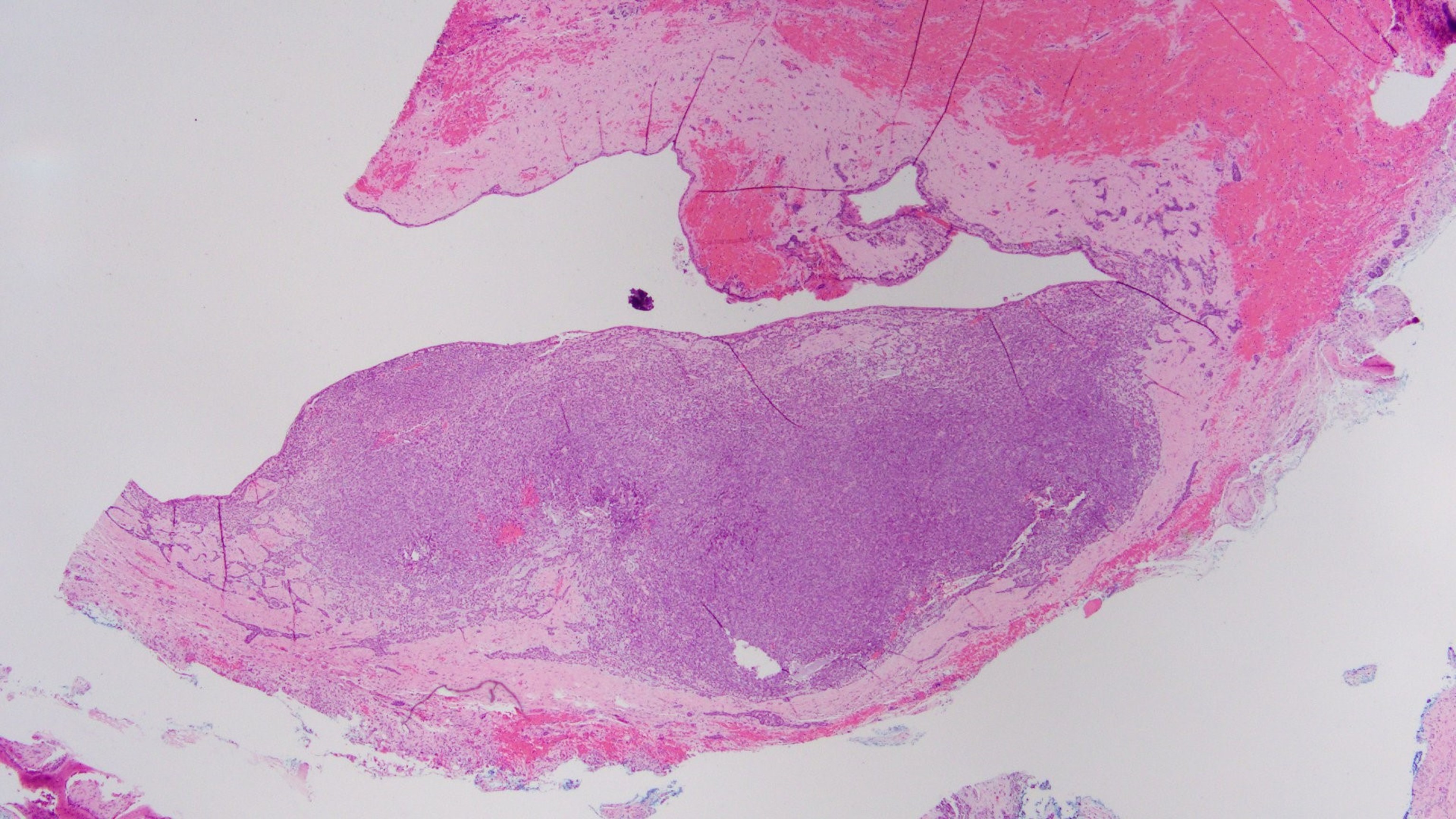
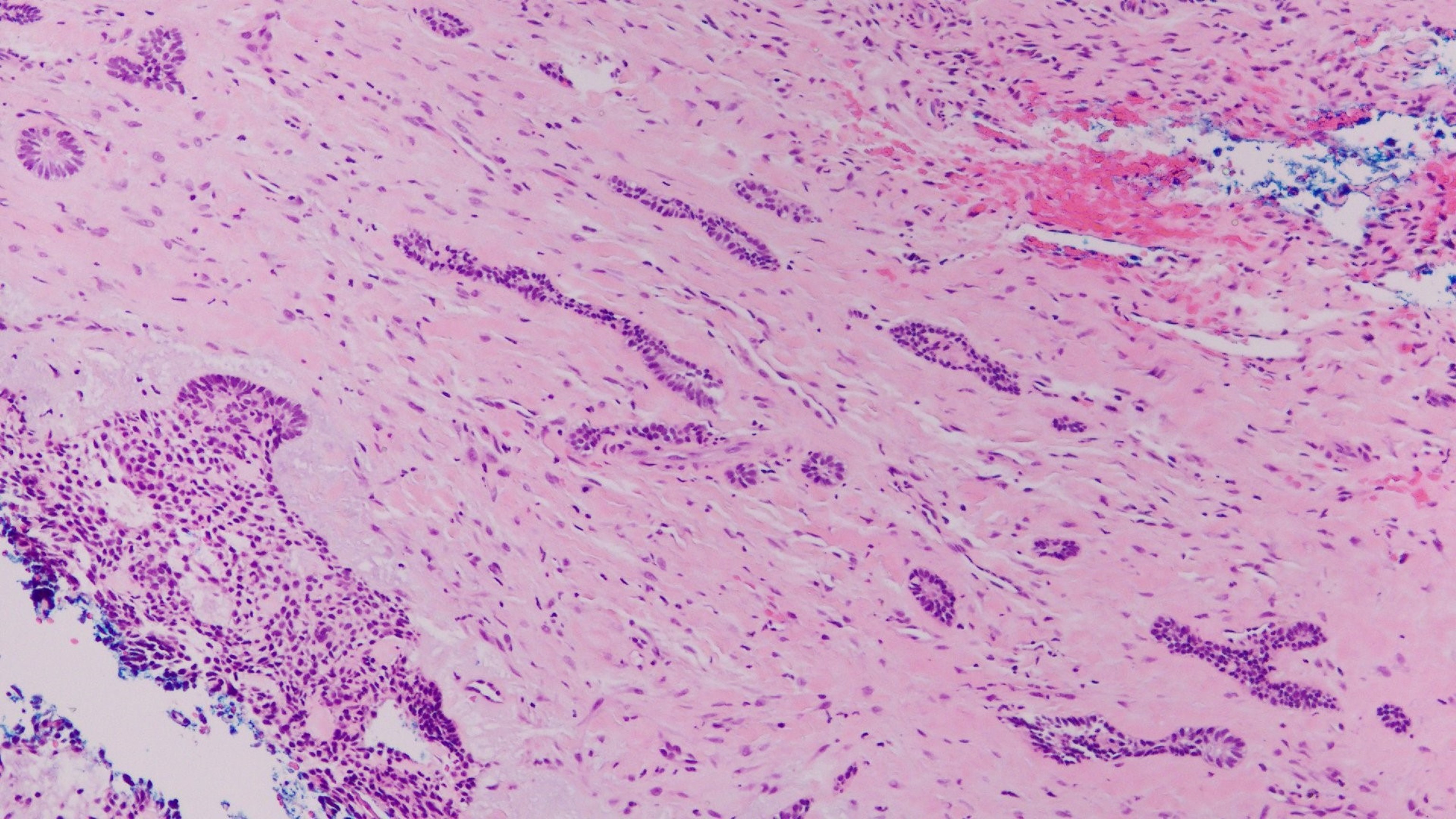
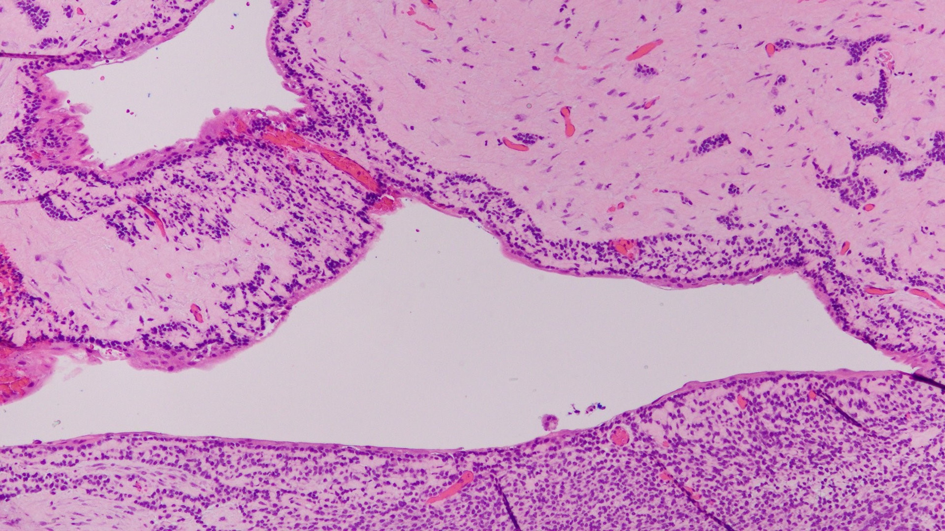
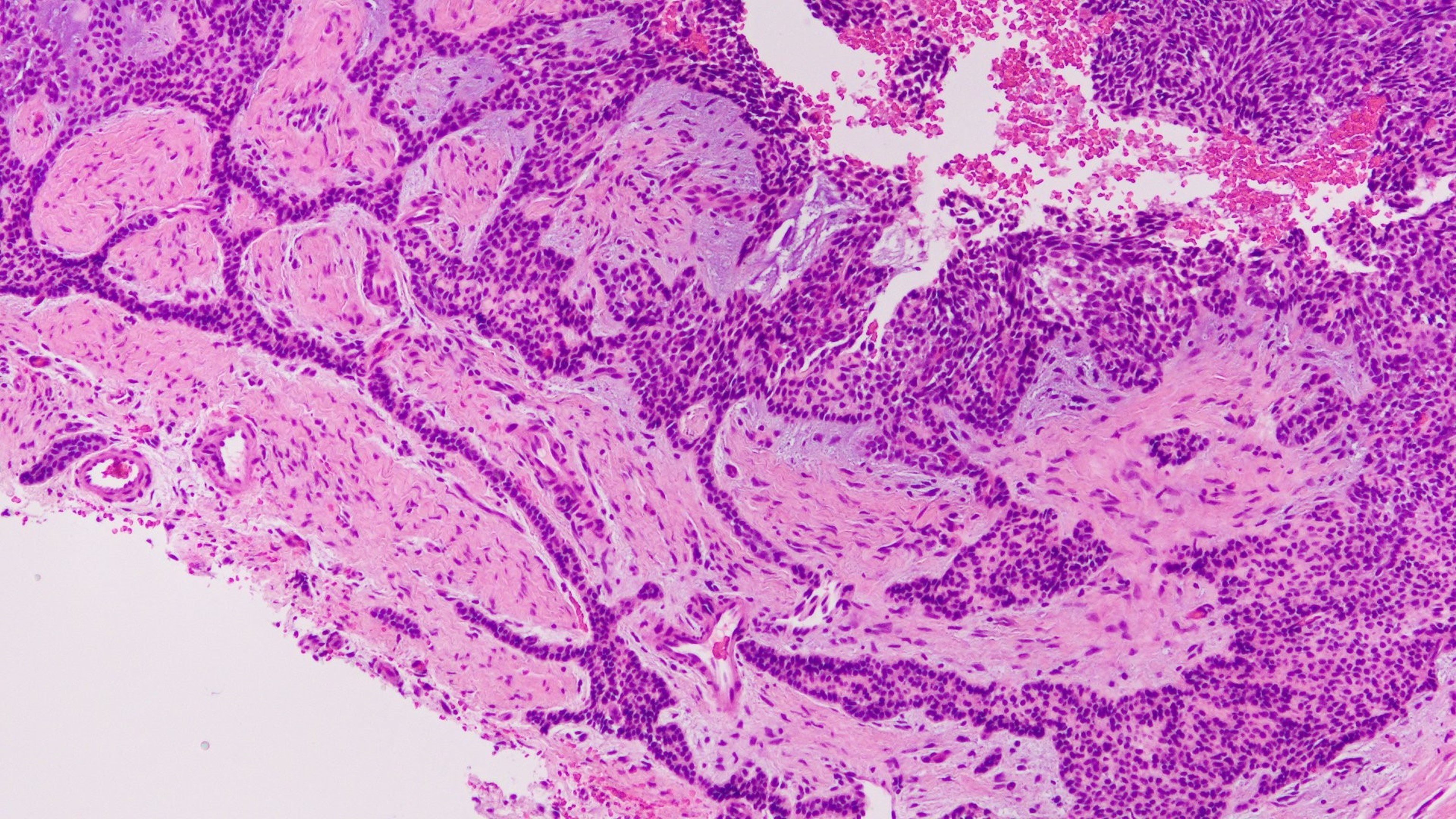
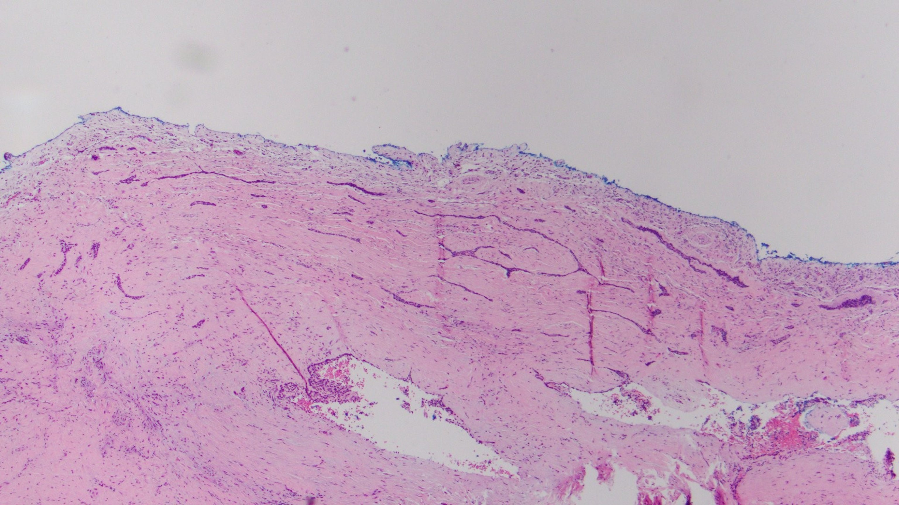
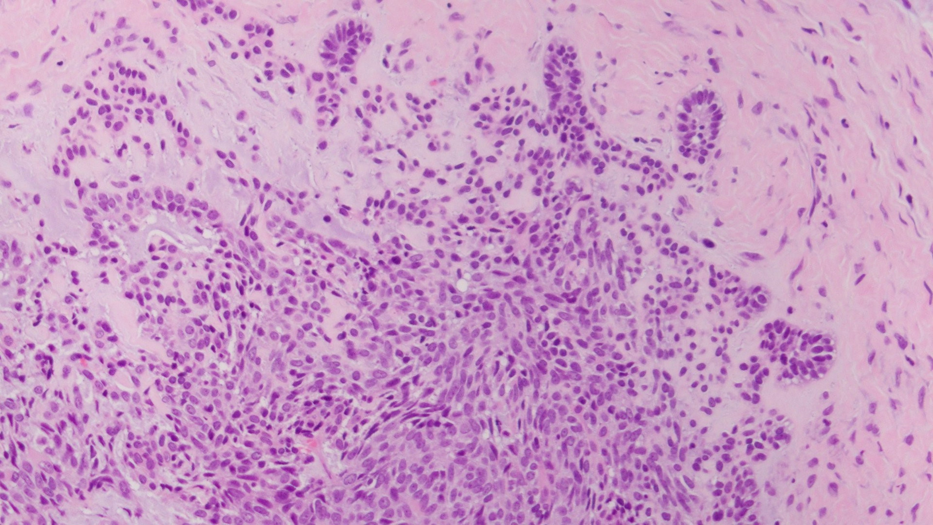
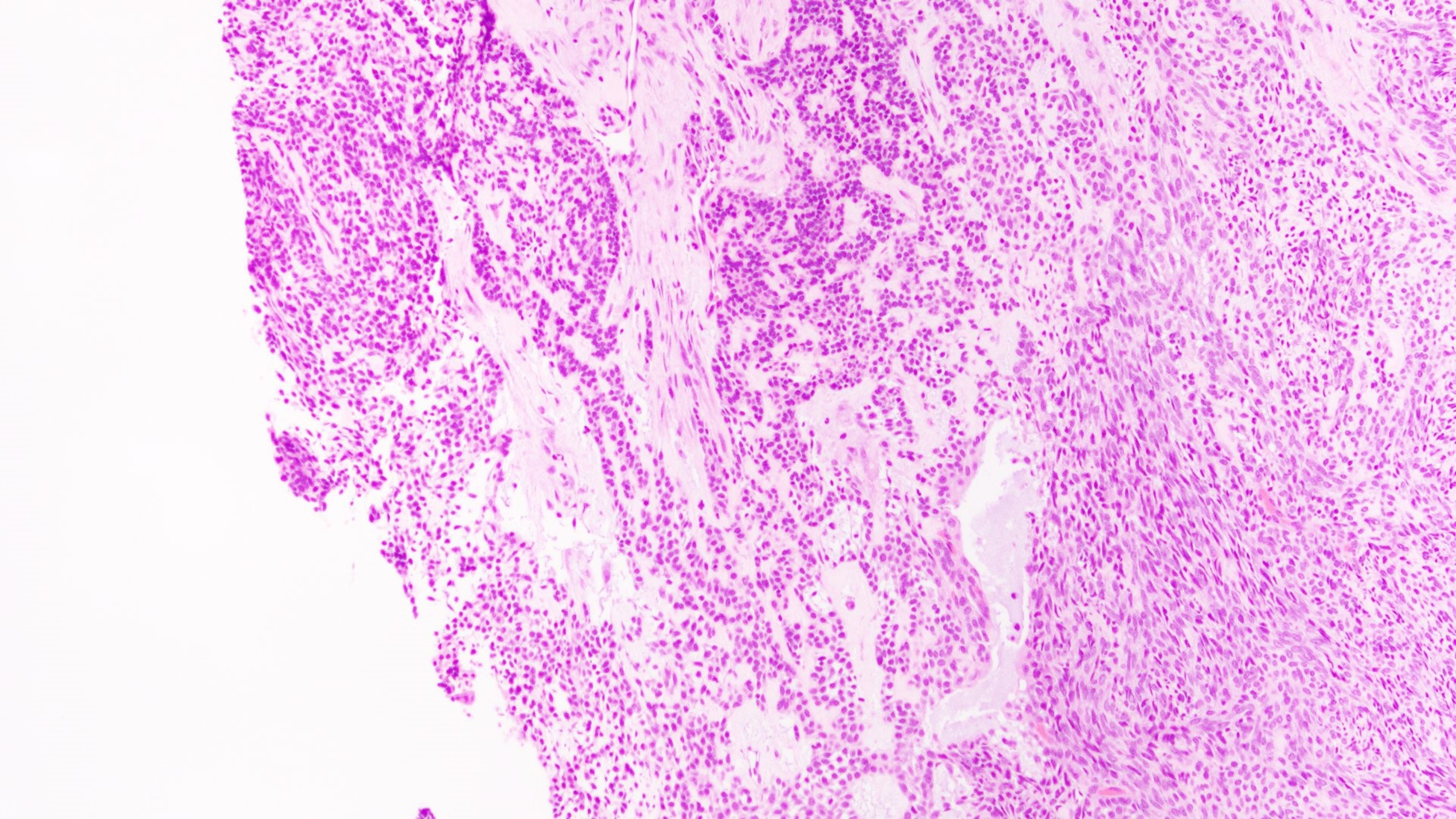
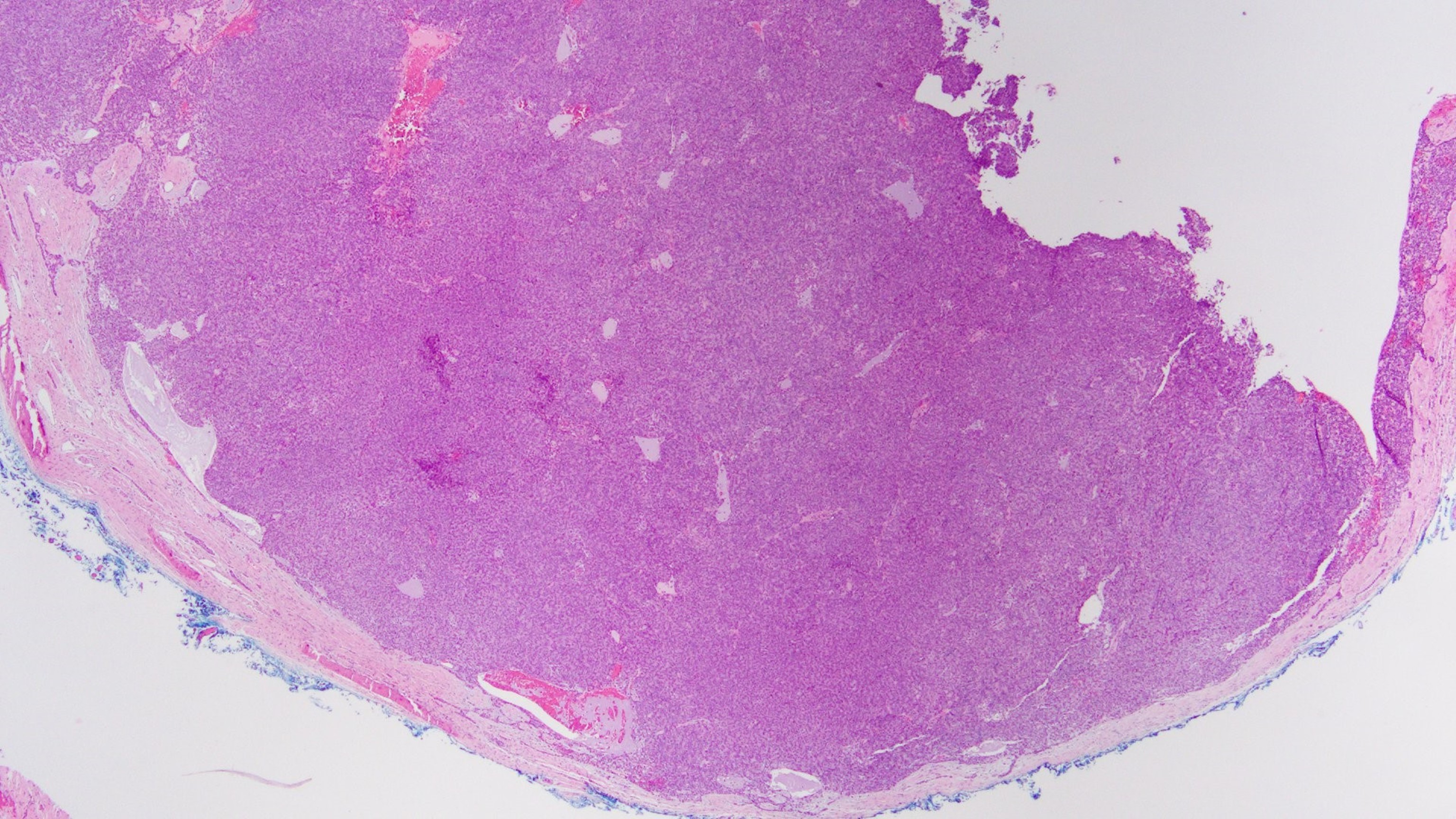
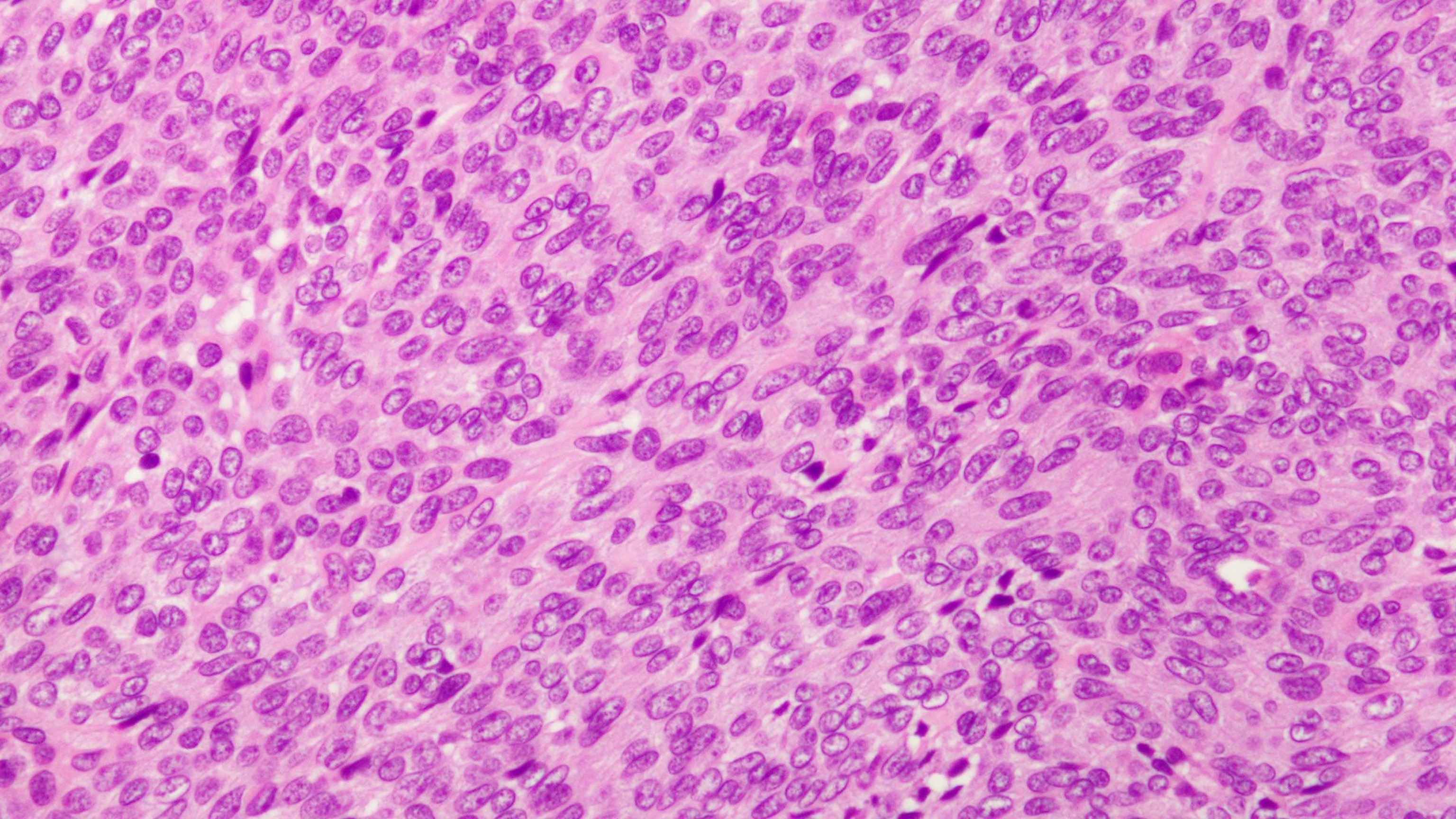
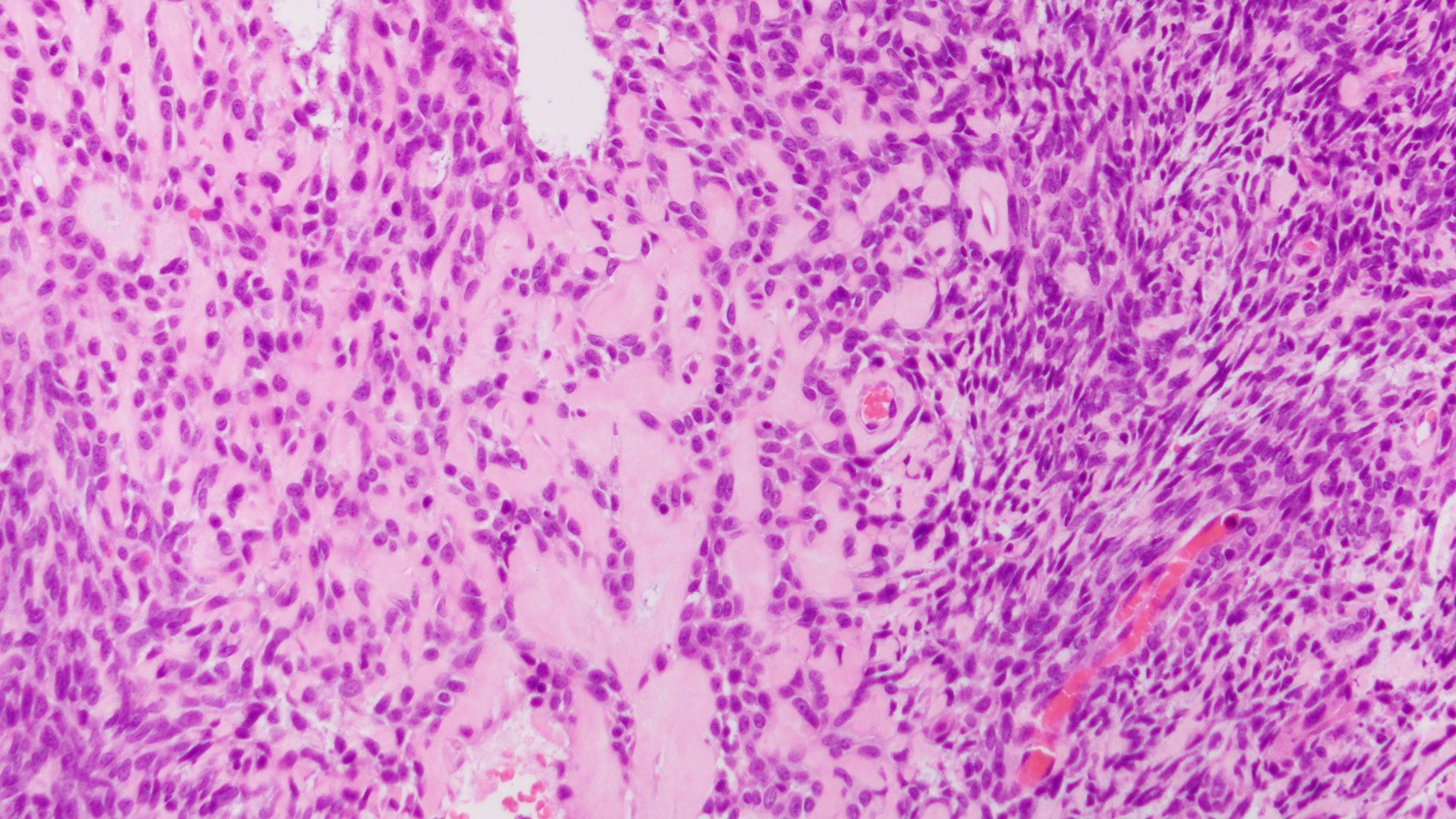
Forum for Clinical and Surgical Oral Pathology Case BBOPF 22-2 Dr. Ivan Stojanov (Case Western Reserve University, School of Dental Medicine, Cleveland, OH. USA) is seeking you input for an unusual case. The case will be posted until February 25, 2022. A summary of the response will ensue shortly after. HistoryA 90M presented to OMFS with a 4.0 cm ant. maxillary RL of unknown duration, exhibiting buccal and palatal expansion but no cortical perforation or erosion (per the surgeon). At time of surgery, a variably solid/cystic mass was encountered with no evidence of encapsulation, but the surgeon claims the lesion was easily removed in its entirety. Received grossly was a ruptured cyst and curettings of soft tissue. Histologic images, attached, show a poorly characterizable odontogenic cystic/neoplastic process. By immunohistochemistry, tumor cells are positive for AE1/3, CK19, p40; and negative for BRAF, beta-catenin (cytoplasmic only), INSM-1. Ki-67 is < 1%. QuestionsHas anyone seen a case similar to this and have a diagnosis? Ivan Stojanov, D.M.D. Images
Case prepared by Dr. Alfredo Aguirre (BBOP Manager) and Daniel Emmer (University at Buffalo School of Dental Medicine). |