Bulletin Board of Oral Pathology
Bulletin Board of Oral Pathology
|
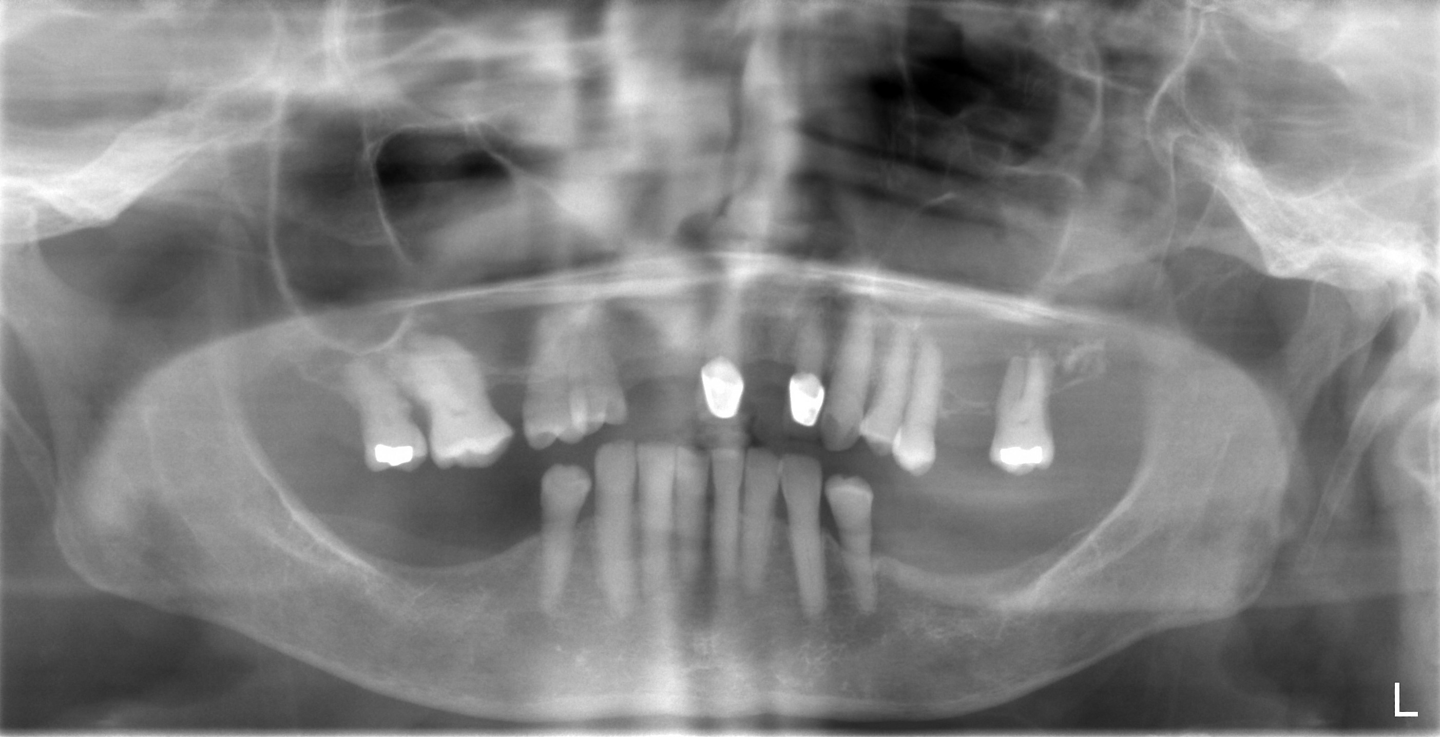
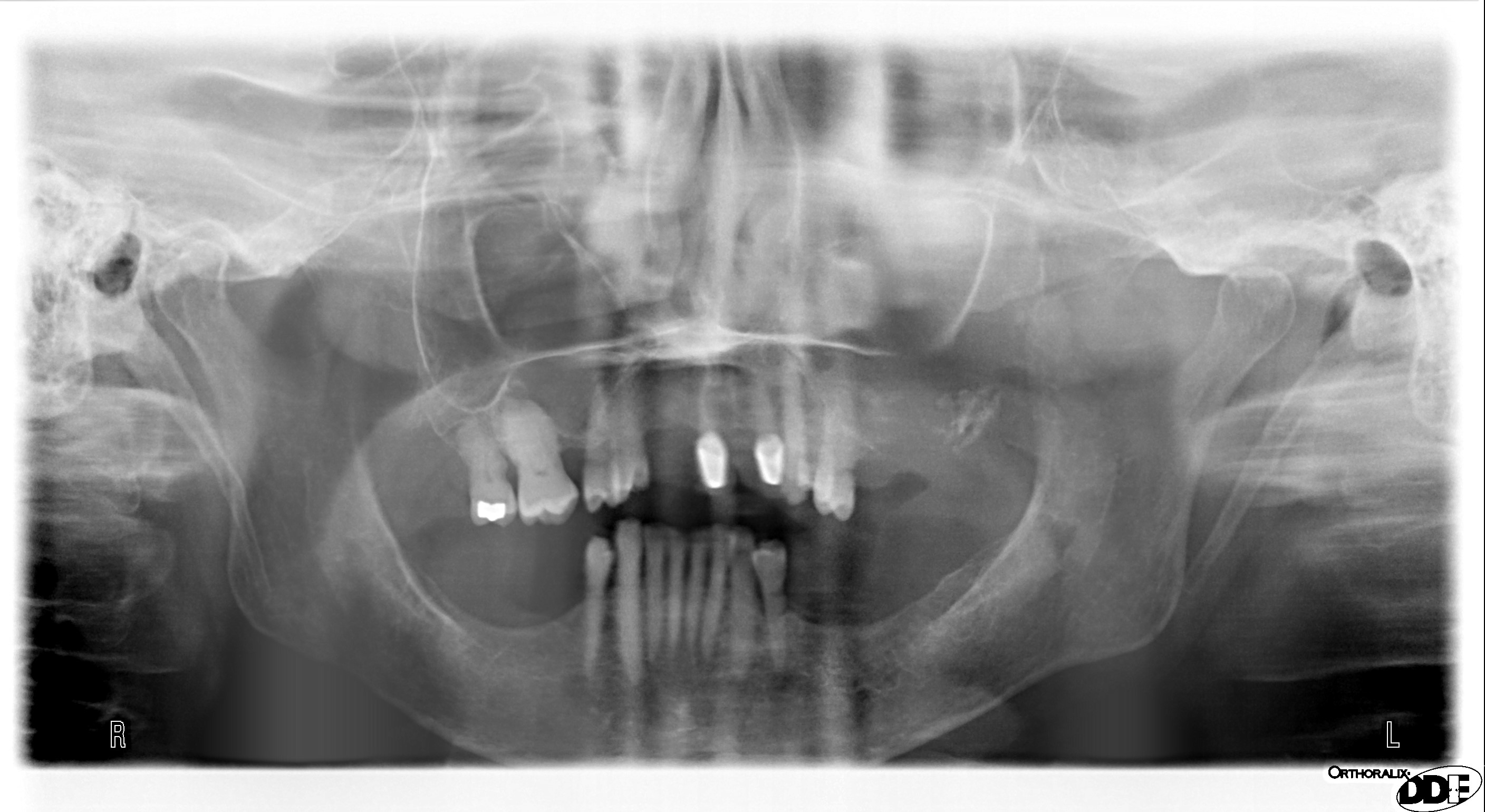
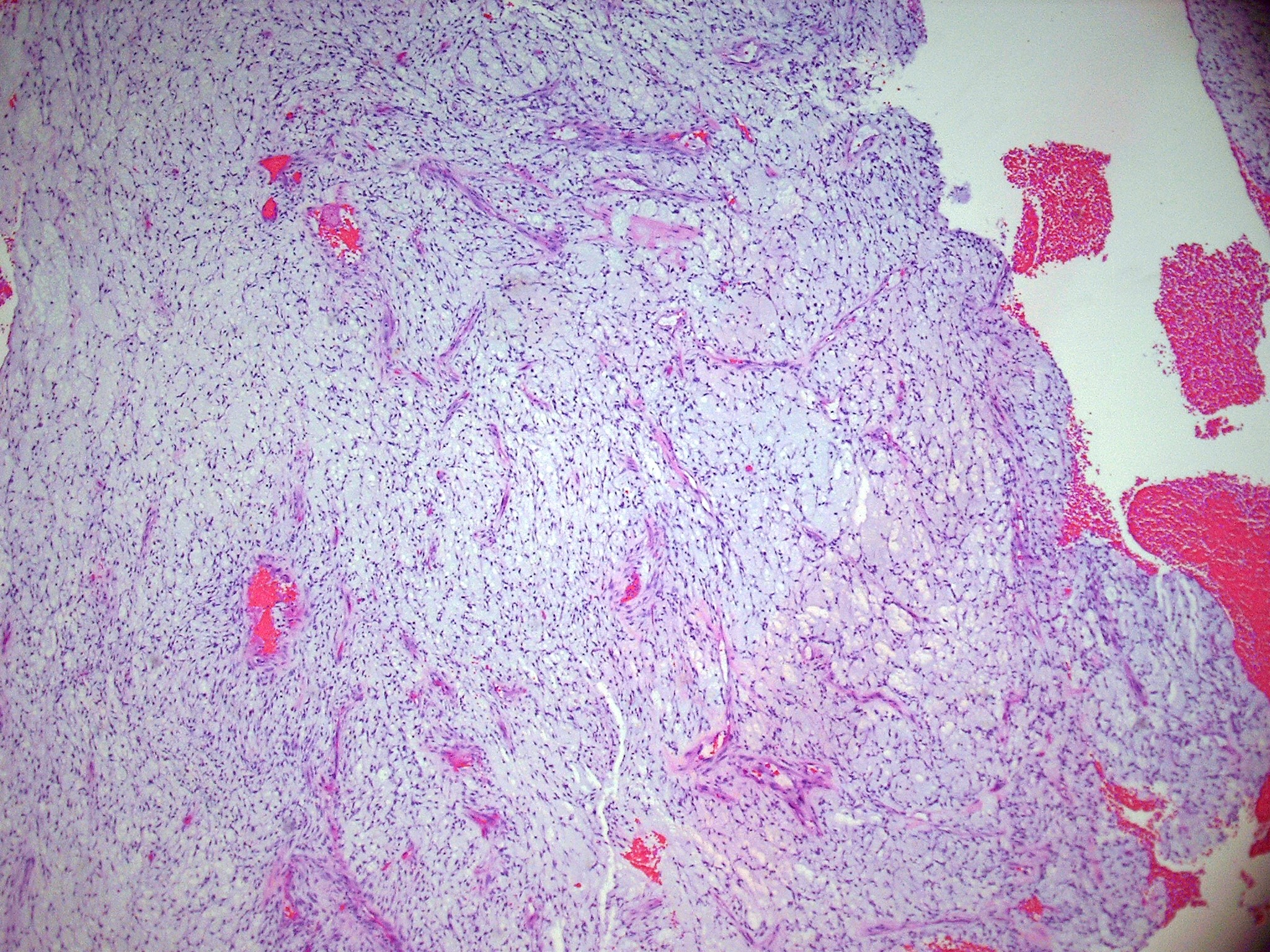
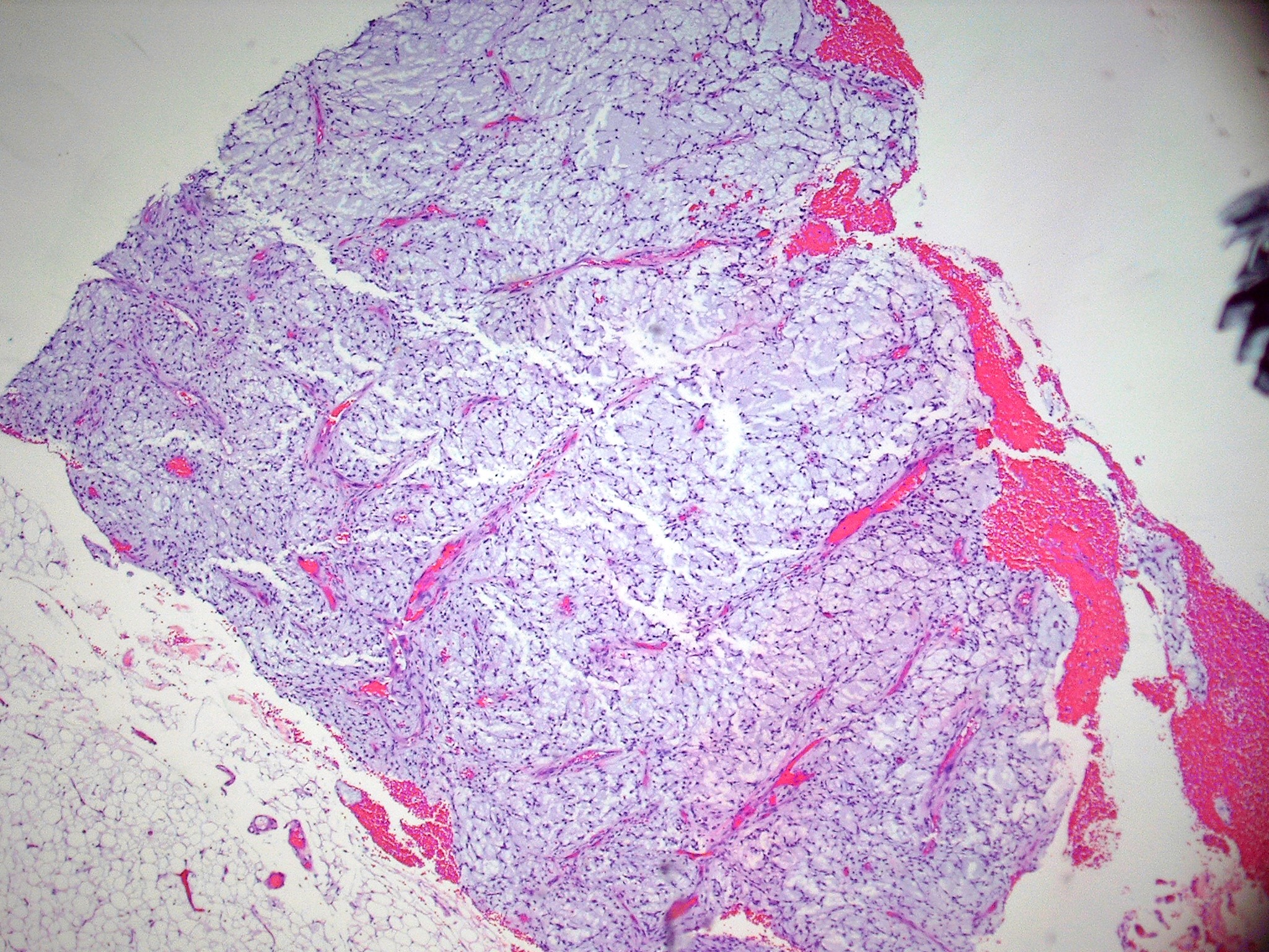
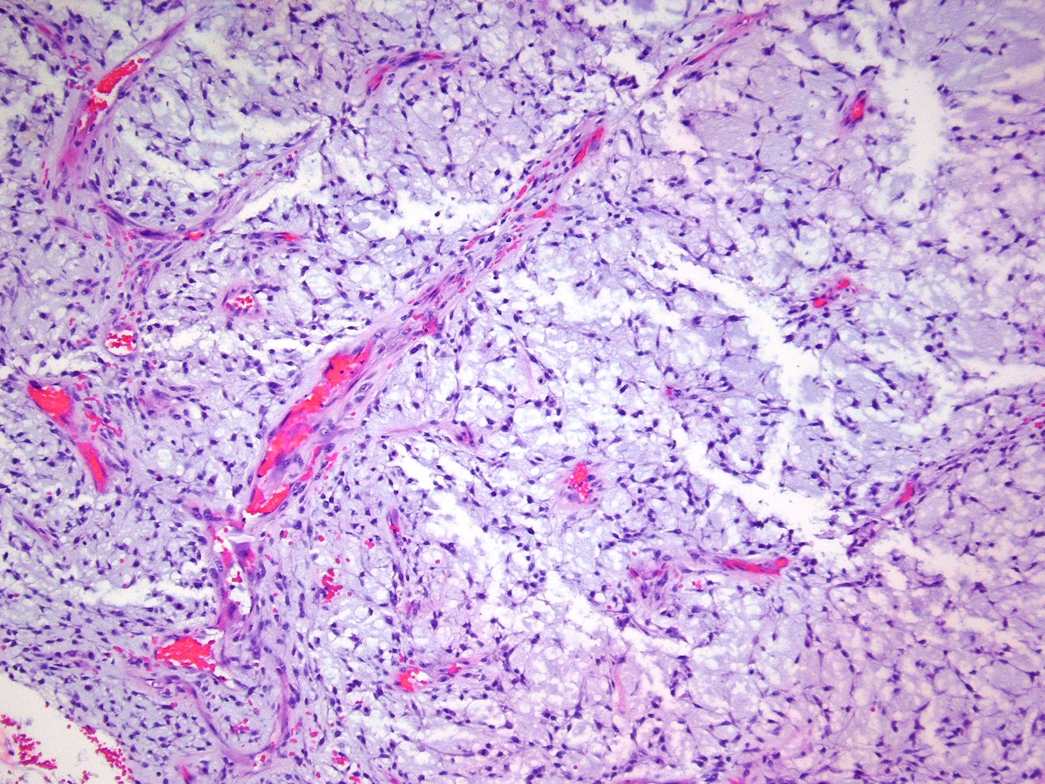
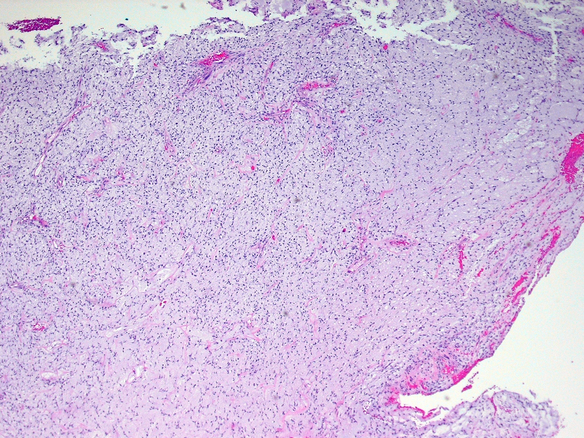
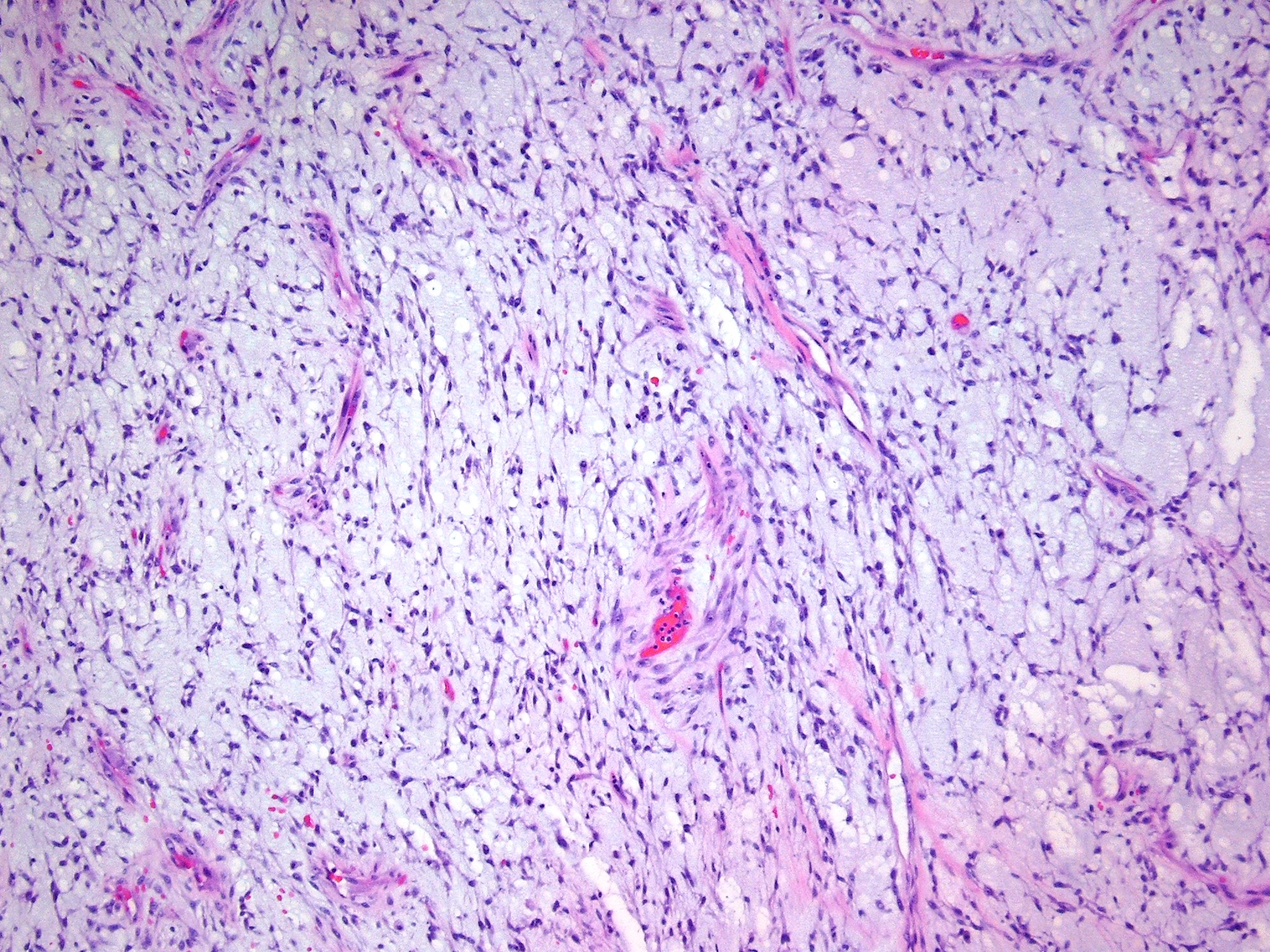
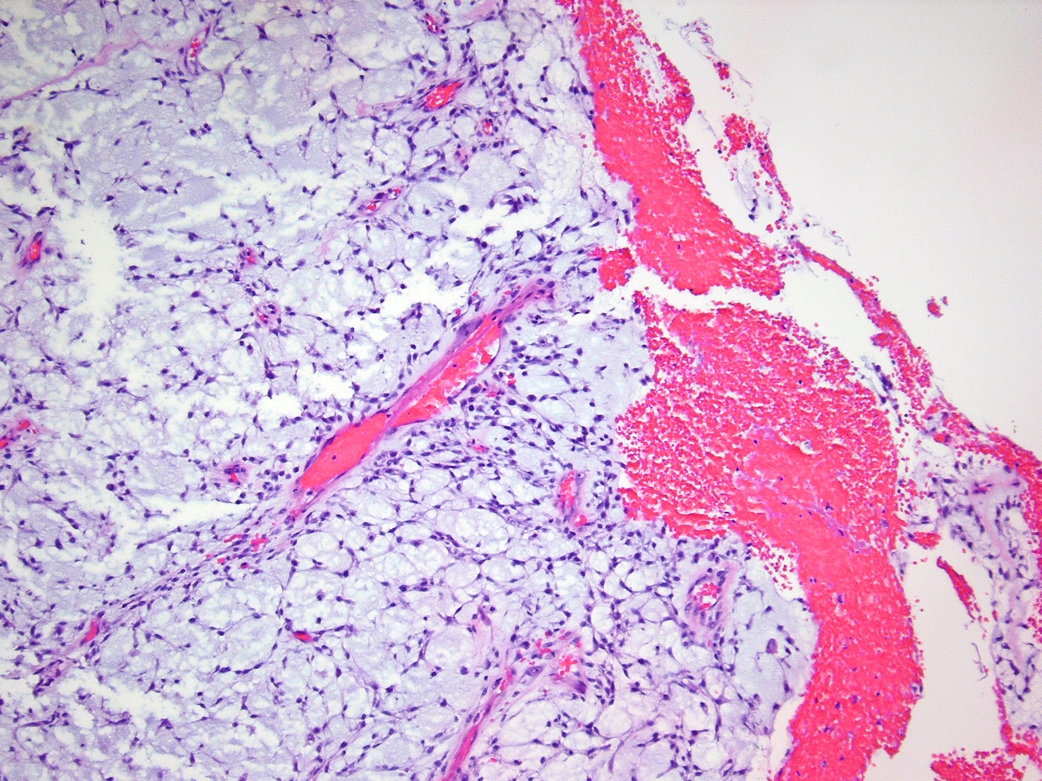
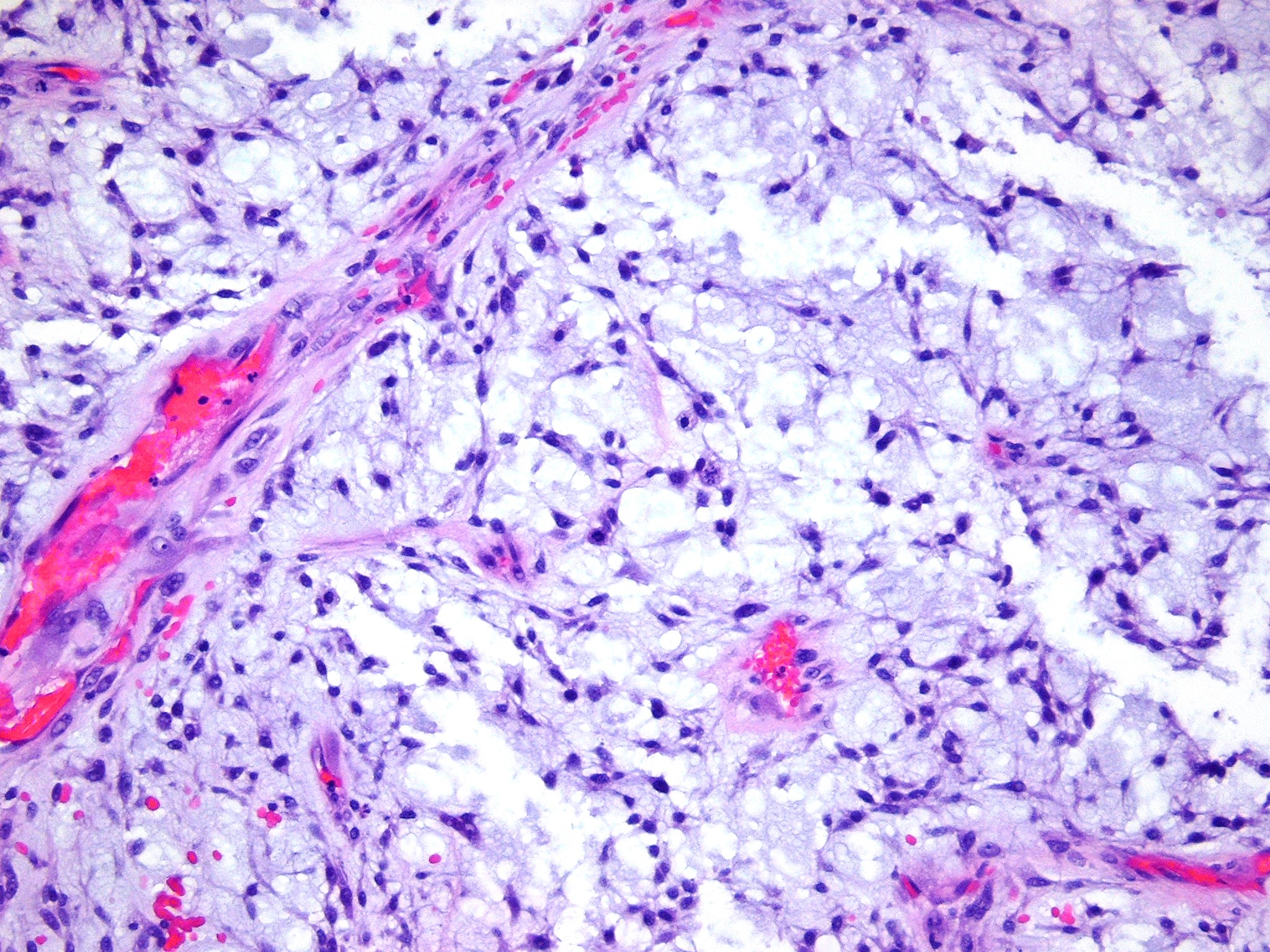
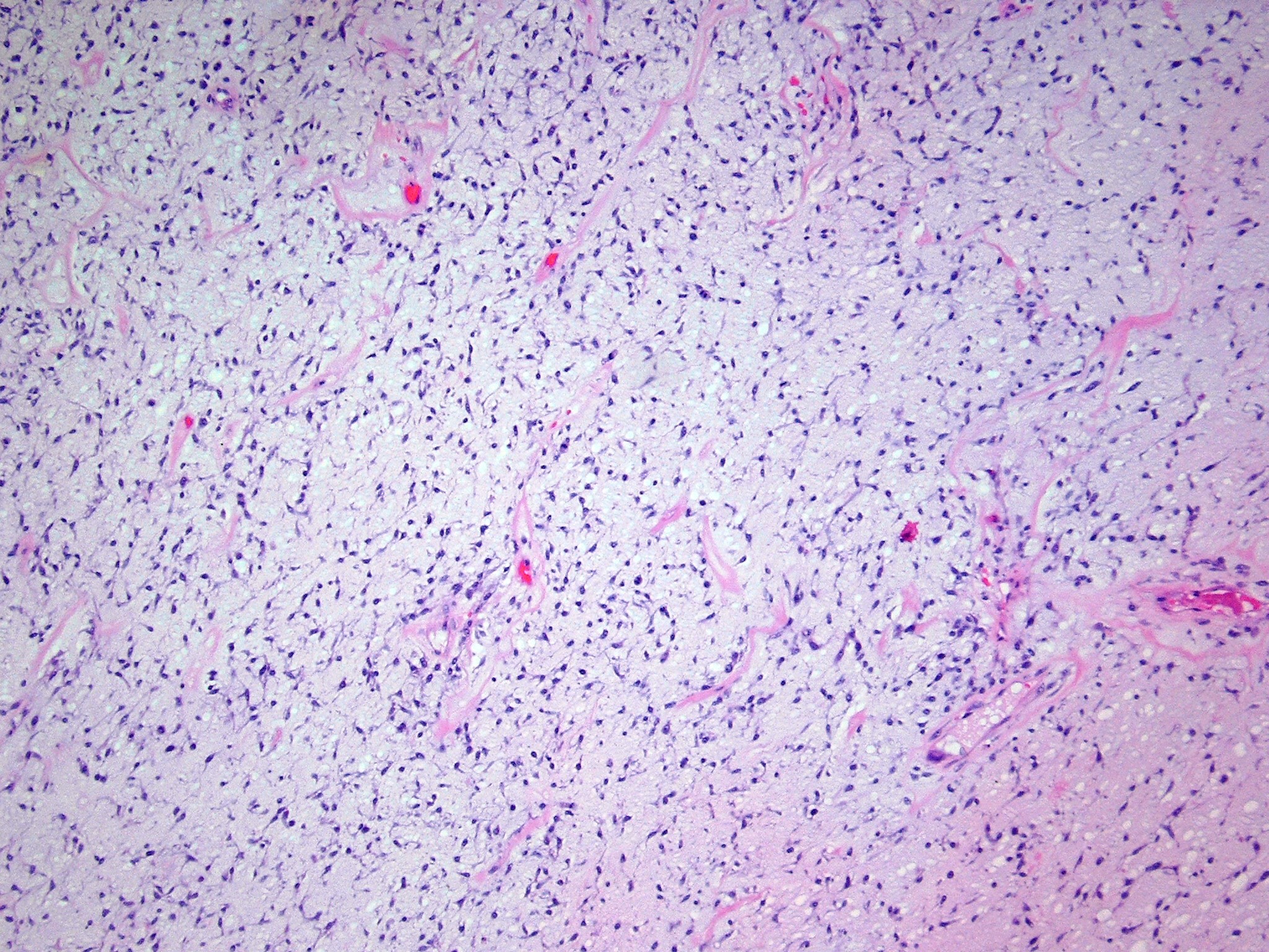
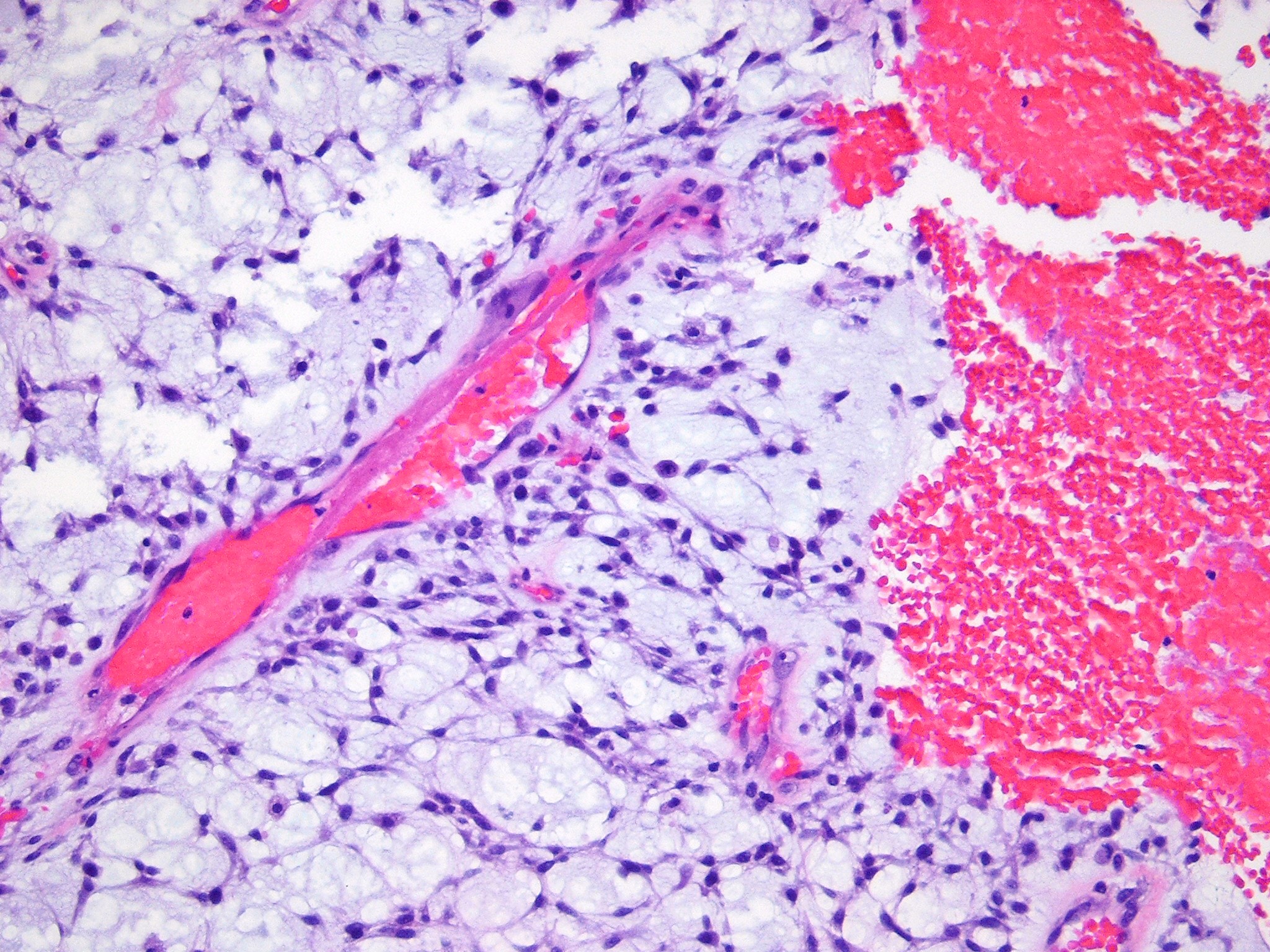
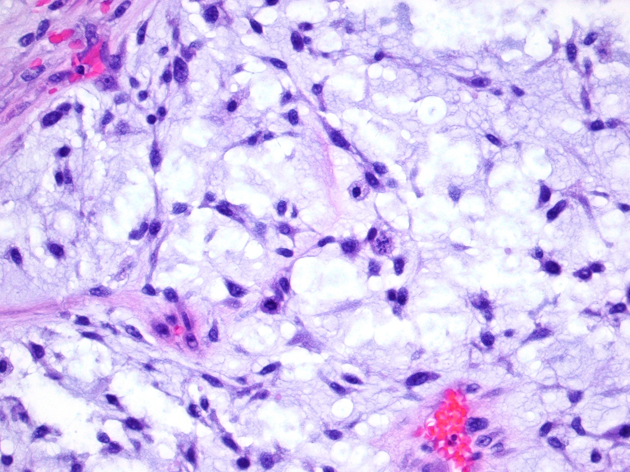
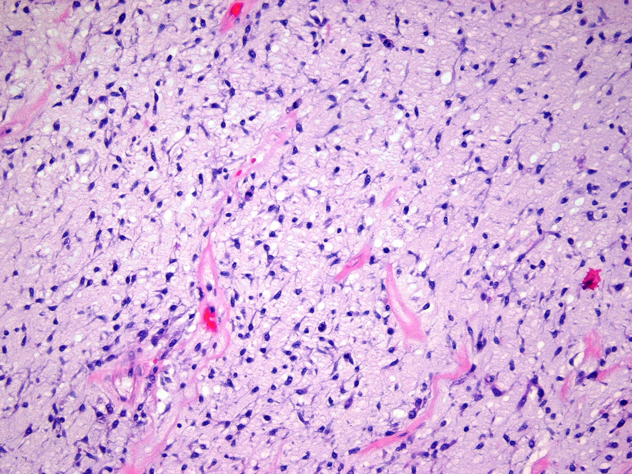
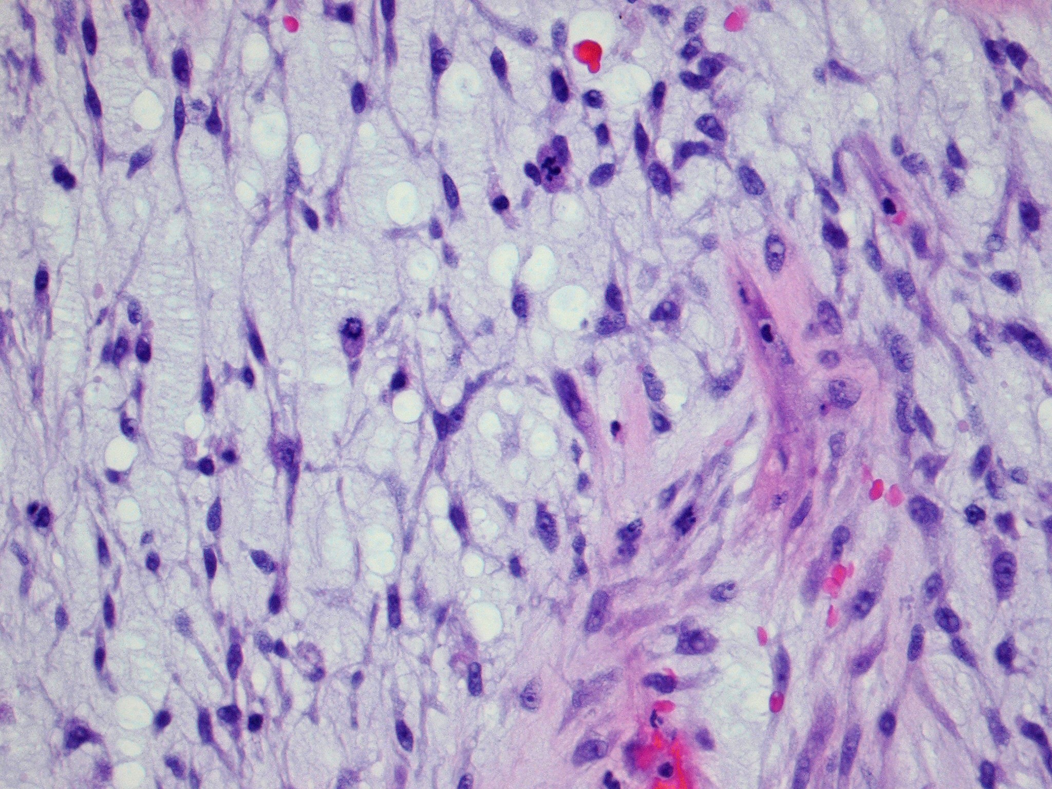
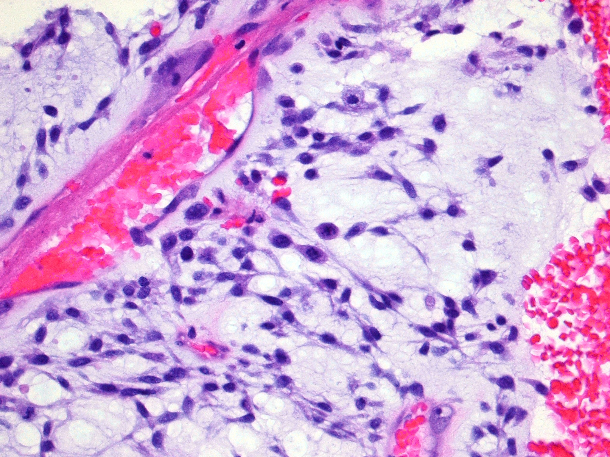
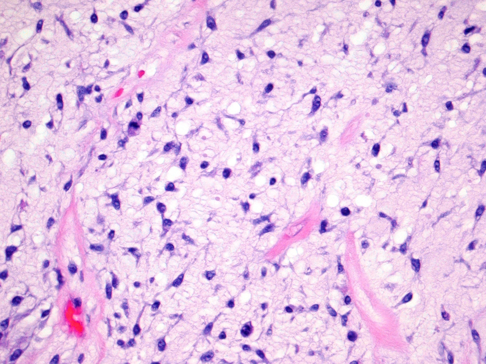
Forum for Clinical and Surgical Oral Pathology Case BBOPF 13-3 This is an 85 year old female who had an upper left molar (#15) tooth extracted. Approximately 3 weeks following the extraction, the patient developed an oral-antral communicating sinus. A surgical procedure was performed to close the fistulous tract. At the time of surgery, the sinus floor was intact but a very large amount of tissue was extracted during the procedure, clinically felt to be a greater volume of tissue than should be expected for the type of procedure performed. I would appreciate your thoughts about this case. Harvey P. Kessler Images(Larger images will open in a new window or tab.)
Case prepared by Dr. Alfredo Aguirre (BBOP Manager) and Daniel Emmer (Web Administrator, University at Buffalo School of Dental Medicine). | ||||||||||||||||||||