Bulletin Board of Oral Pathology
Bulletin Board of Oral Pathology
|
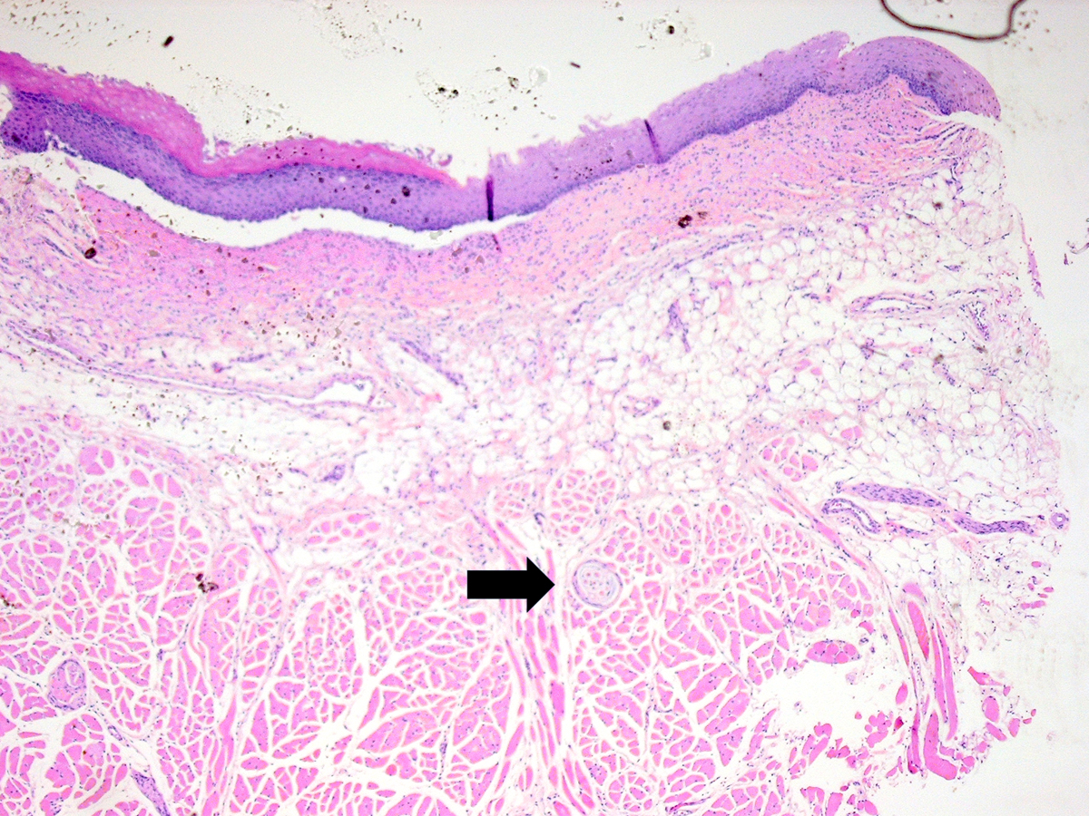
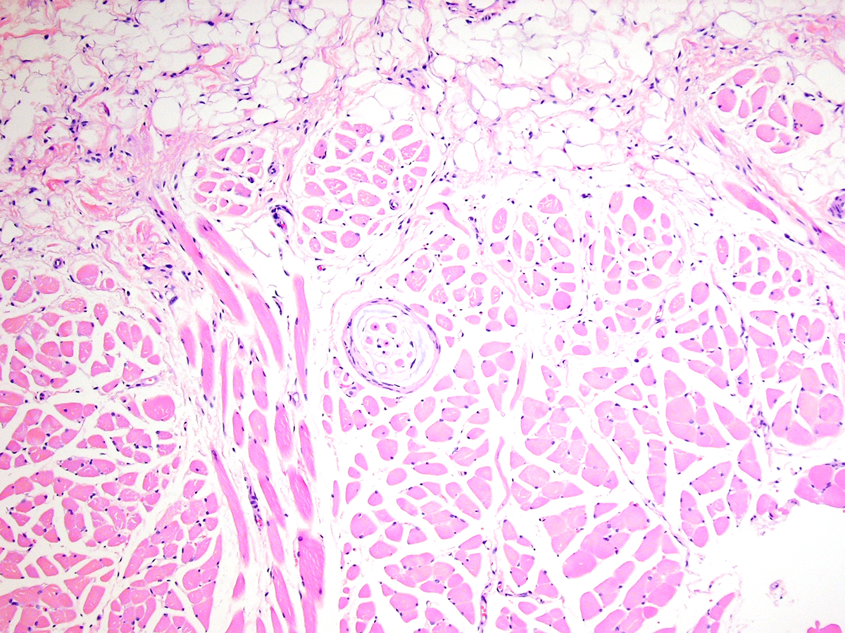
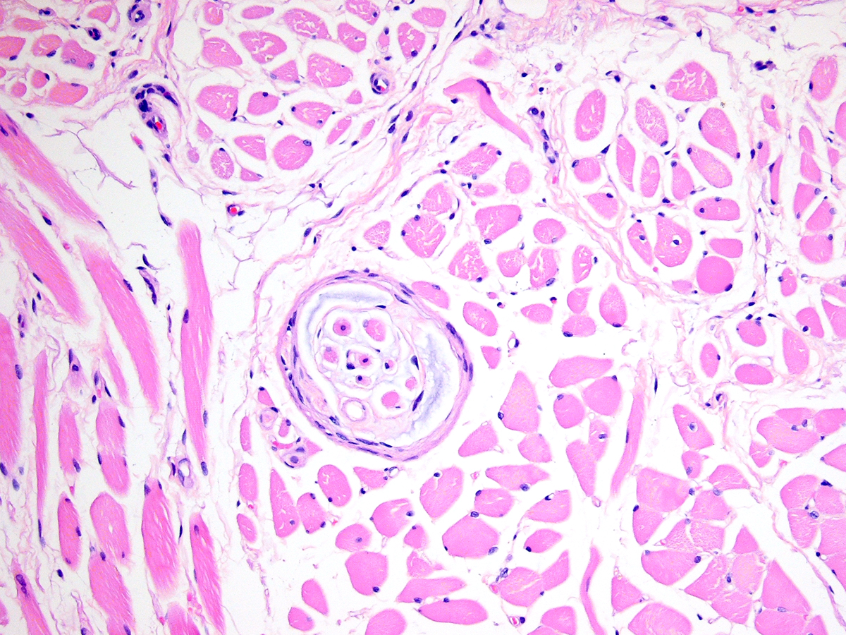
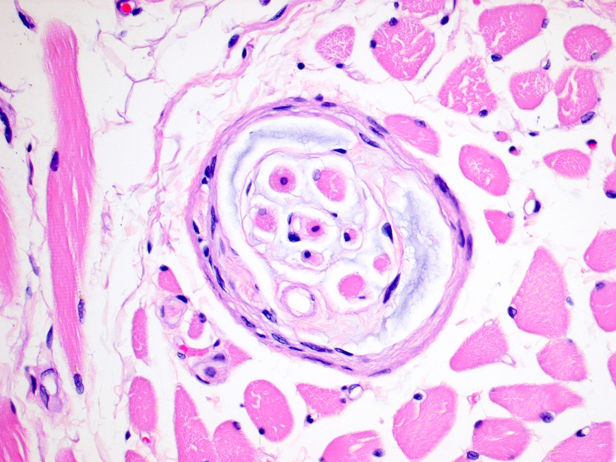
Forum for Clinical and Surgical Oral Pathology Case BBOPF 11-2 Dr. Harvey Kessler (Baylor College of Dentistry. Dallas, Texas. USA) is requesting your opinion. Please send your comments in the window located below the images. This case will be posted from October 14-21, 2011. A summary of the responses will be posted in BBOP. NarrativeI ran across an interesting small structure in a tongue biopsy as an incidental finding and I don’t know for sure what it is. I am posting the following images to ask if anyone knows for sure what it might be. There are 4 photomics at 2x, 5x, 10x, and 20x. There is an arrow on the 2x photomicrograph for the purpose of orientation and location of the structure relative to the muscle layers and the overlying mucosa. From low power it appeared to be a nerve with ganglion cells but the "ganglion cells" clearly are muscle fibers when you go down on them and compare them to the adjacent skeletal muscle fibers. I wondered if this might be a very fortunate section through an area of the motor/neural endplate where a nerve inserts on the muscle surface, but I don’t really appreciate any nerve fibers within the round structure. Does anyone recognize this material with certainty? Dr. Harvey Kessler Images(Larger images will open in a new window or tab.)
Case prepared by Dr. Alfredo Aguirre (BBOP Manager) and Daniel Emmer (Web Administrator, University at Buffalo School of Dental Medicine). |