Bulletin Board of Oral Pathology
Bulletin Board of Oral Pathology
|
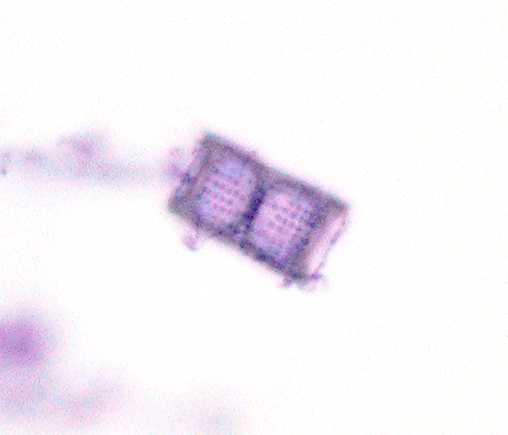
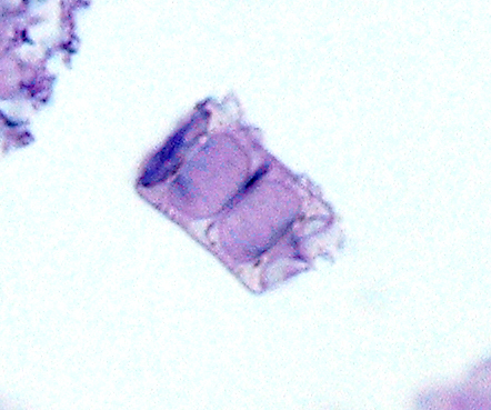
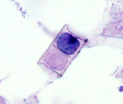
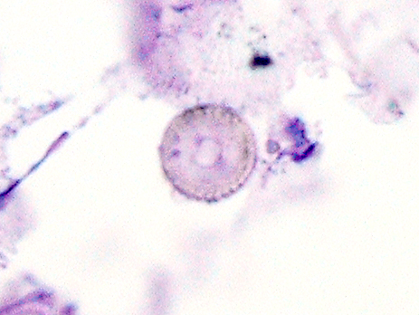
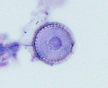
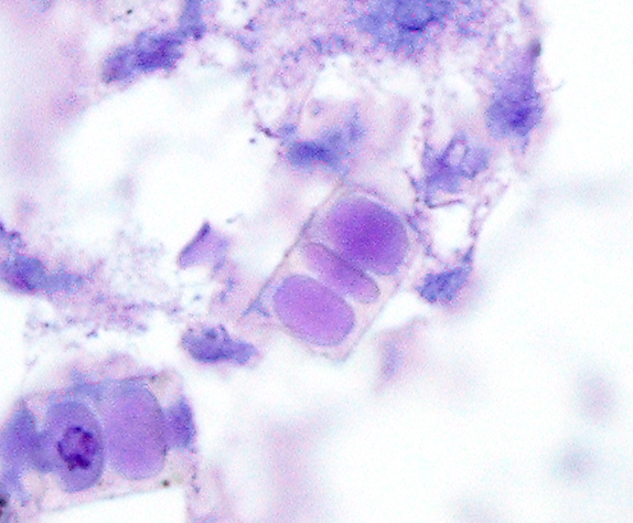
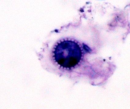
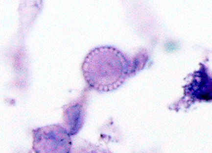
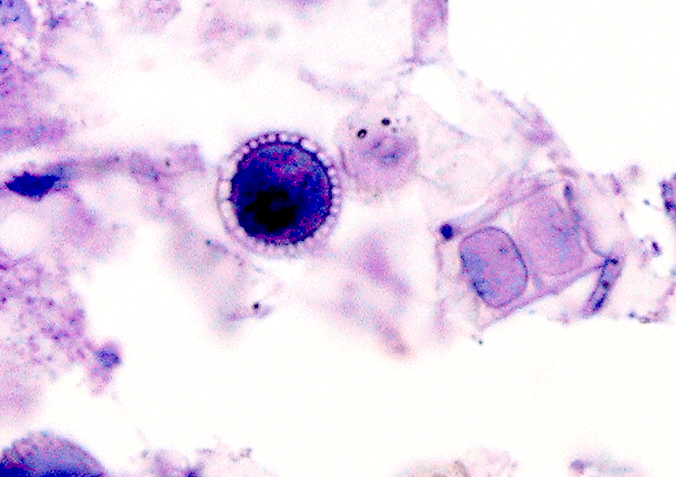
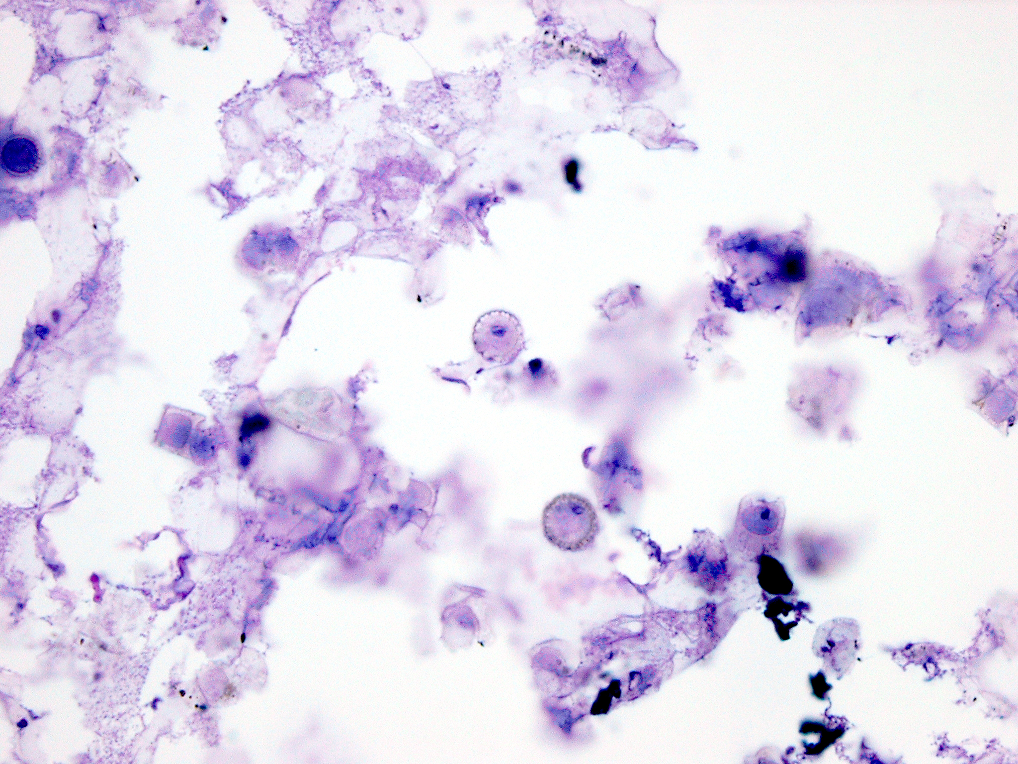
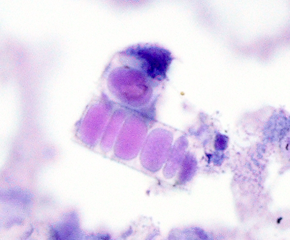
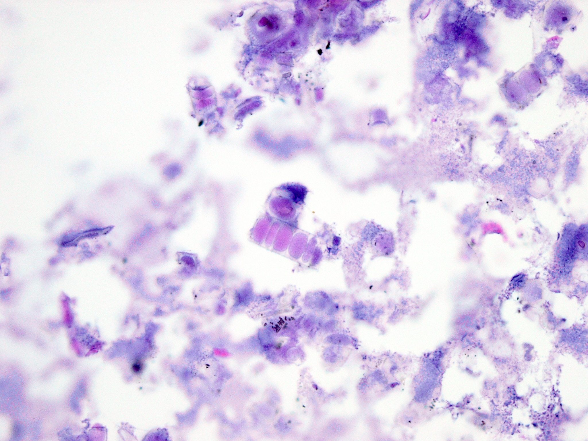
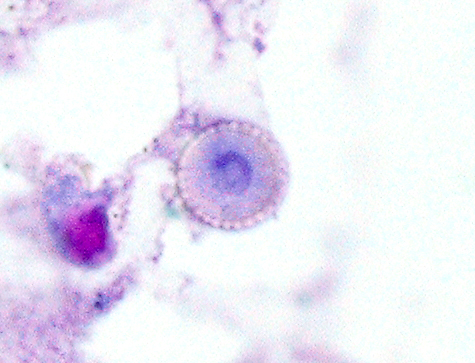
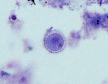
Forum for Clinical and Surgical Oral Pathology Case BBOPF 10-2 Dr. Harvey Kessler (Baylor College of Dentistry. Dallas, Texas. USA) is requesting your opinion. Please send your comments in the window located below the images. This case will be posted from April 8-15, 2010. A summary of the responses will be posted in BBOP. Clinical History60 year-old white female. Specimen removed from the maxillary sinus. That's all we know. At gross examination, the material was the consistency of inspissated sinus mucin--very loose, jelly-like, gray color, slightly opaque or cloudy appearing. I wondered if it would even survive the processing, but it did. There isn't anything but the mucoid material in the specimen--no soft tissue components. Scattered throughout though, are two very distinctive structures (D10-1846-9 and -11 and -13). One looks like a flower petal but the central portion is bigger and the "petals" are small, almost like a cuticle (D10-1846-10 and -14 and -15). Some of these show a hint of pigment in the "petal" area (D10-1846-4). The central area in some of them stains pretty dark (D10-1846-7); in most though, the center stains weakly (D10-1846-8). There is a round structure that stains lighter within the central portion in many of them (D10-1846-5). The 2nd pattern looks like a cinderblock (D10-1846-2 and -3). Most of these have the classic cinderblock pattern of two roughly square "holes" in the center, but occasional ones are more elongated and have more "holes." (D10-1846-6 and -12) If you focus up and down thru the thickness of some of the cinderblocks, an occasional one will show a lattice or waffle-like texture (D10-1846-1) The images were taken with oil at 60x so some are a little fuzzy--please excuse this, but most of the material is thick enough that it is tough to get it in a single focal plane. I've cropped and resized many of the images to try to highlight the materials. We don't recognize this as a fungal organism and believe it is some type of foreign material, possibly pollen. It does not polarize however. We showed it to a general path colleague who is our resident weird bug expert, and he also doesn't think it is a fungal organism but is not entirely confident. Does anyone recognize this material with certainty? Dr. Harvey Kessler Images(Larger images will open in a new window or tab.) Case prepared by Dr. Alfredo Aguirre (BBOP Manager) and Daniel Emmer (Web Administrator, University at Buffalo School of Dental Medicine). |