Bulletin Board of Oral Pathology
Bulletin Board of Oral Pathology
|
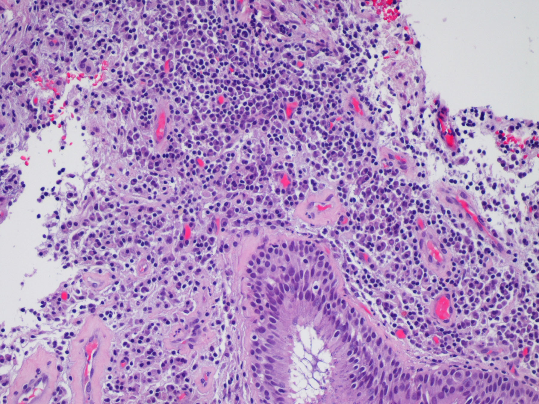
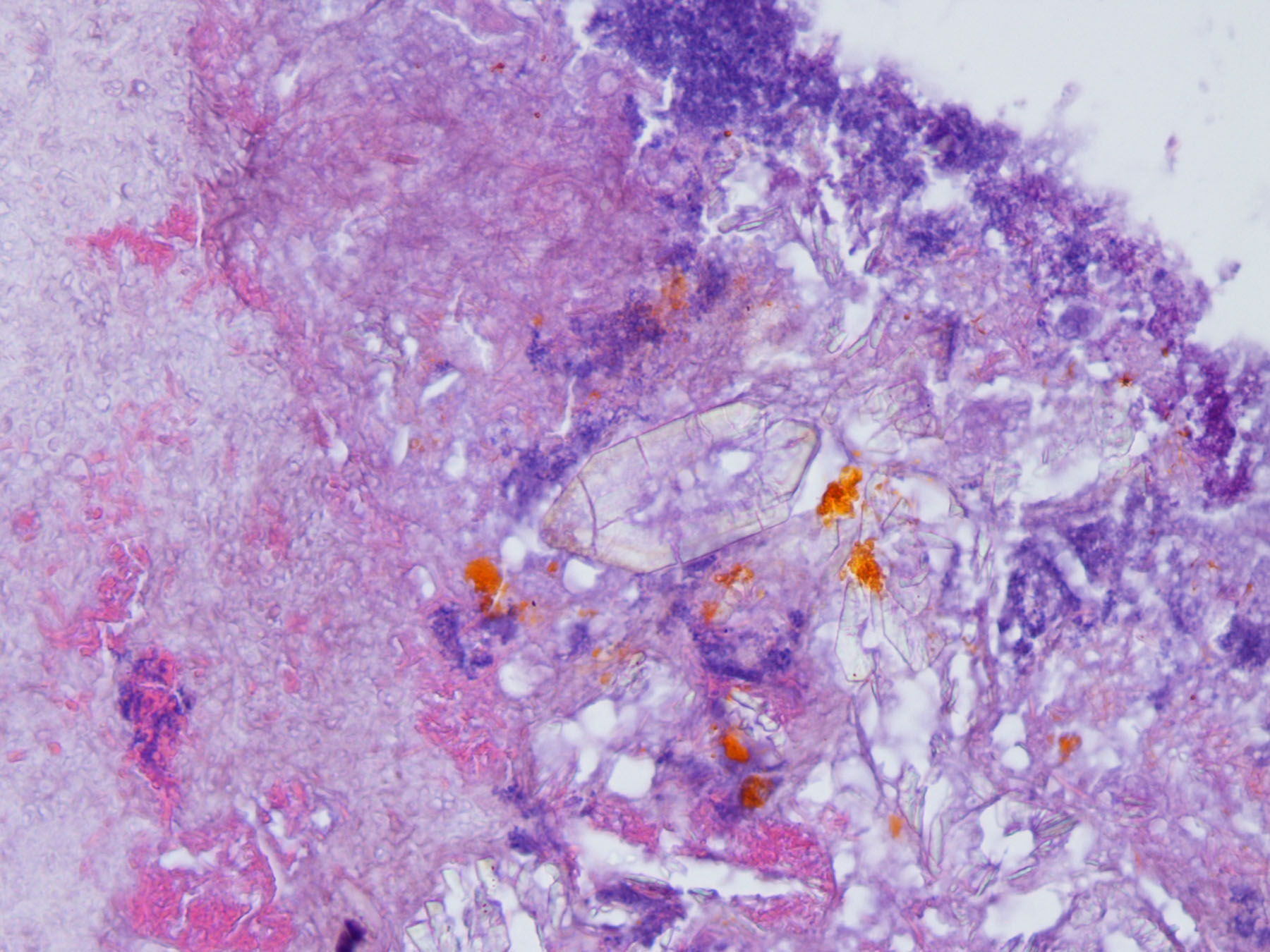
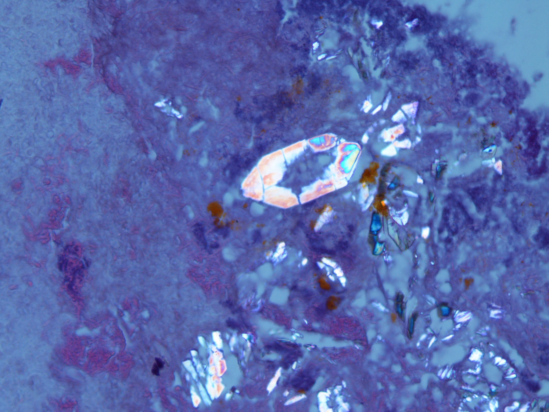
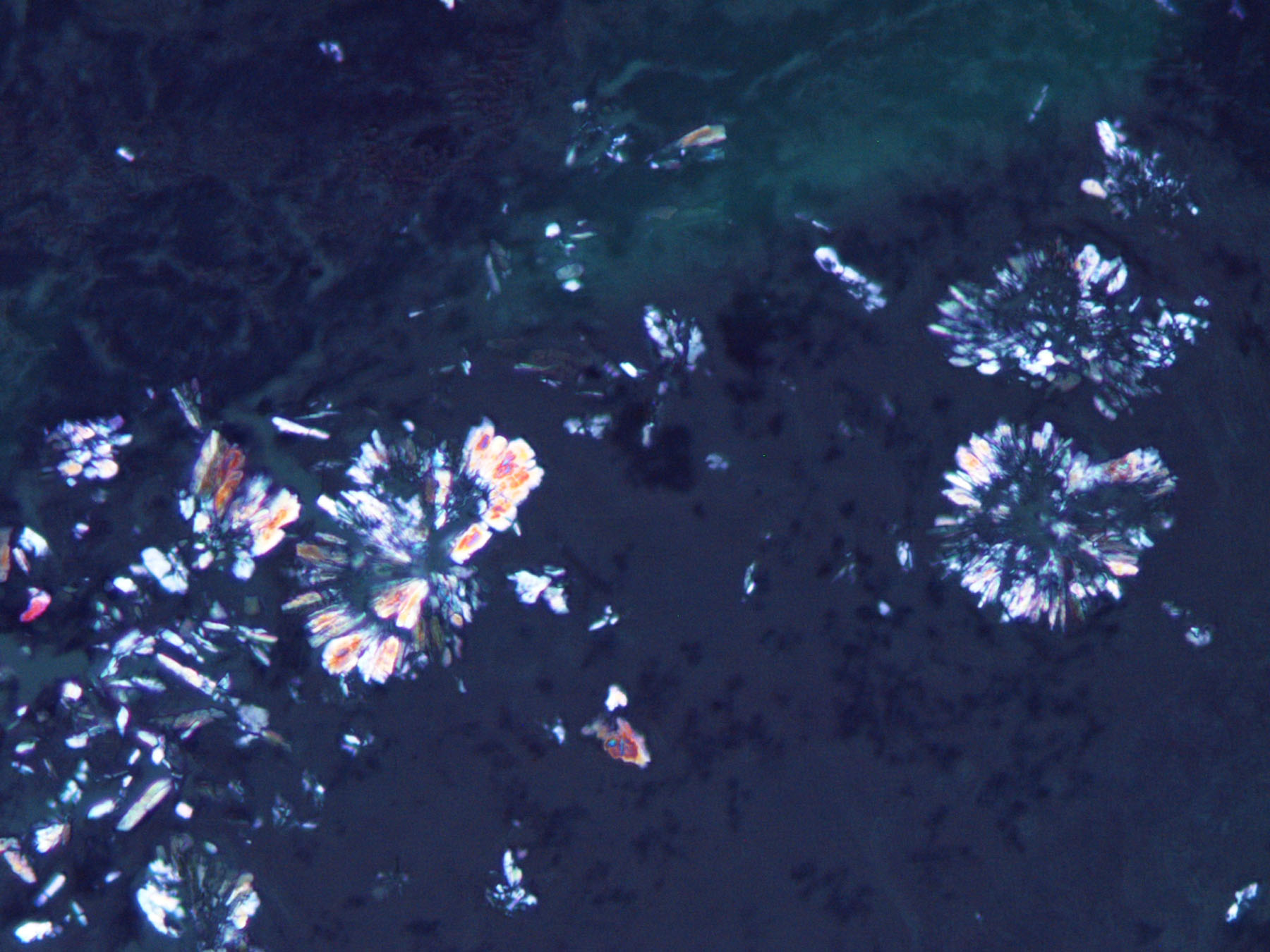
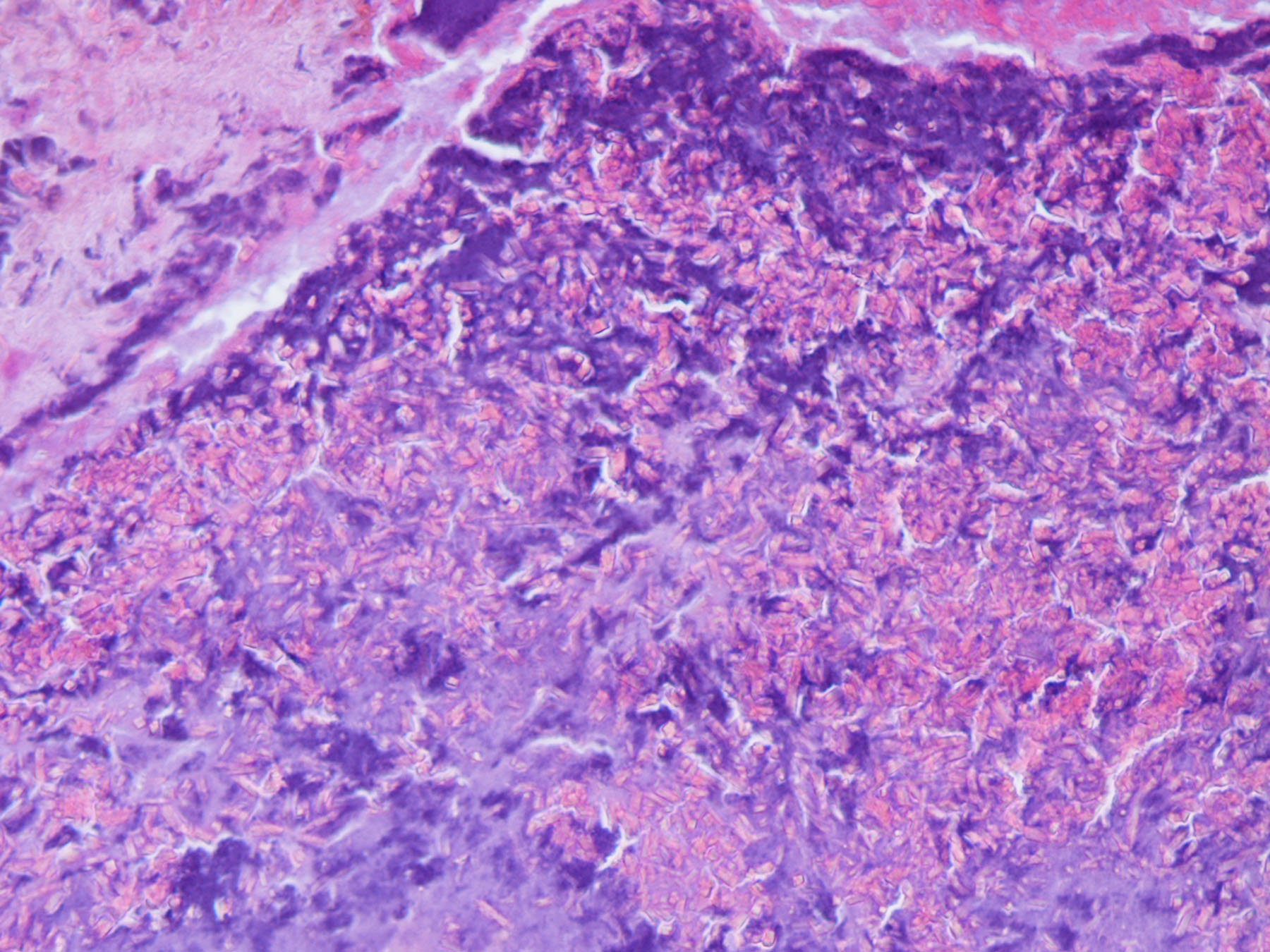
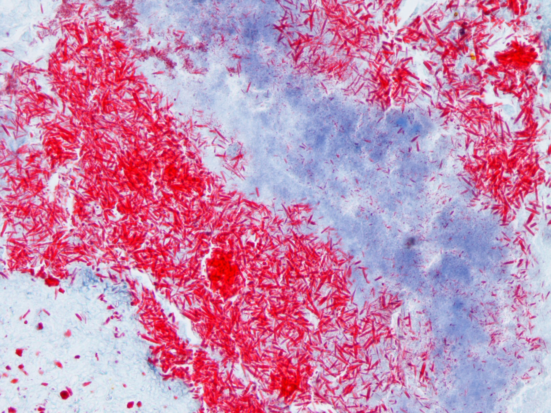
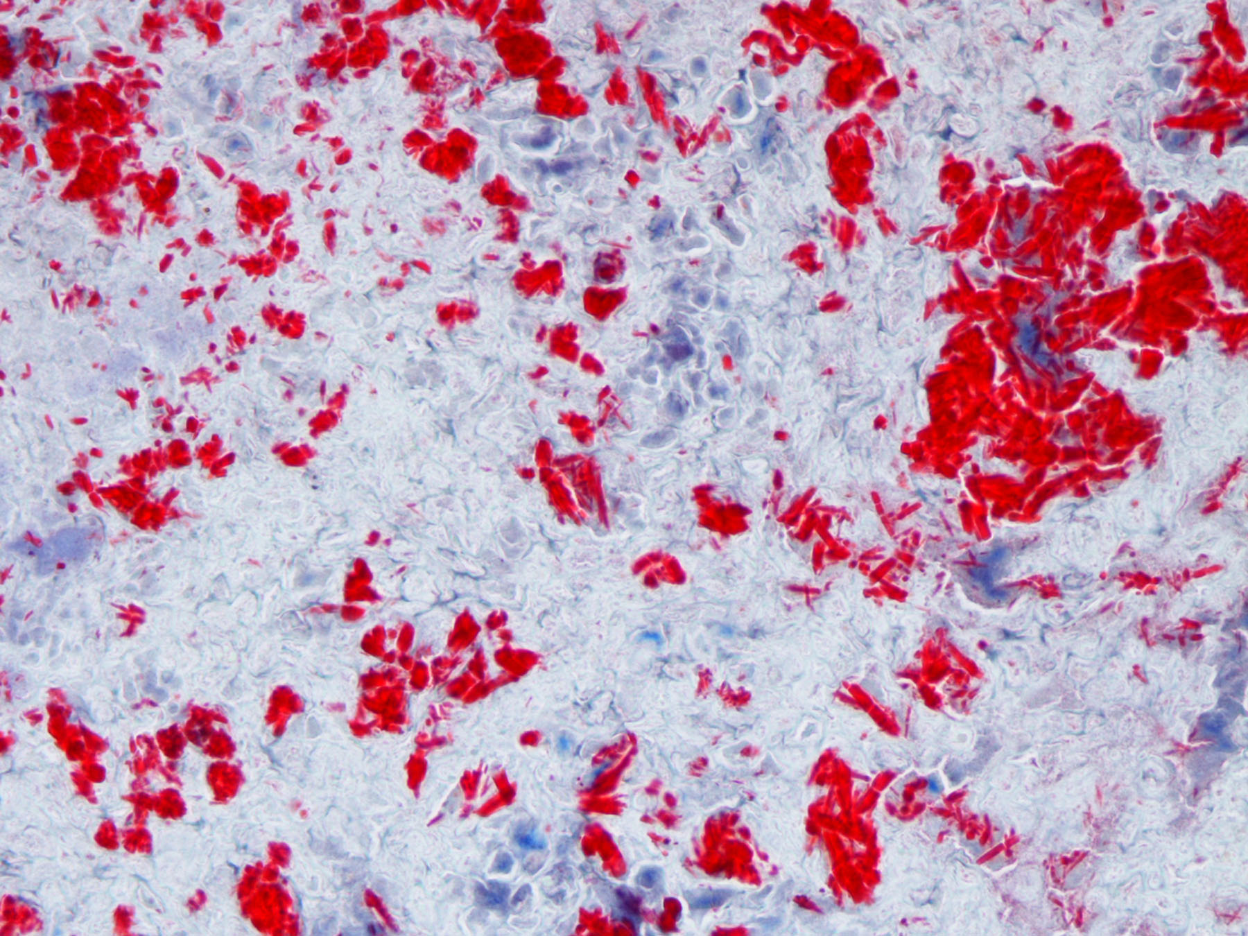
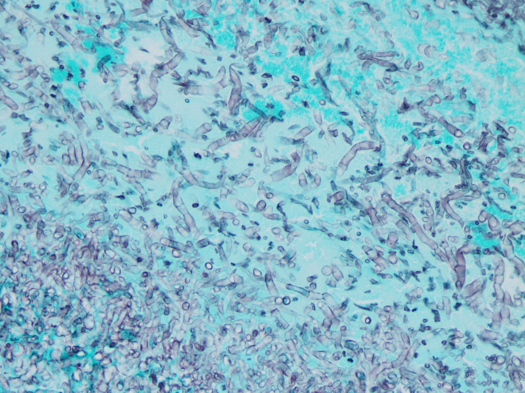
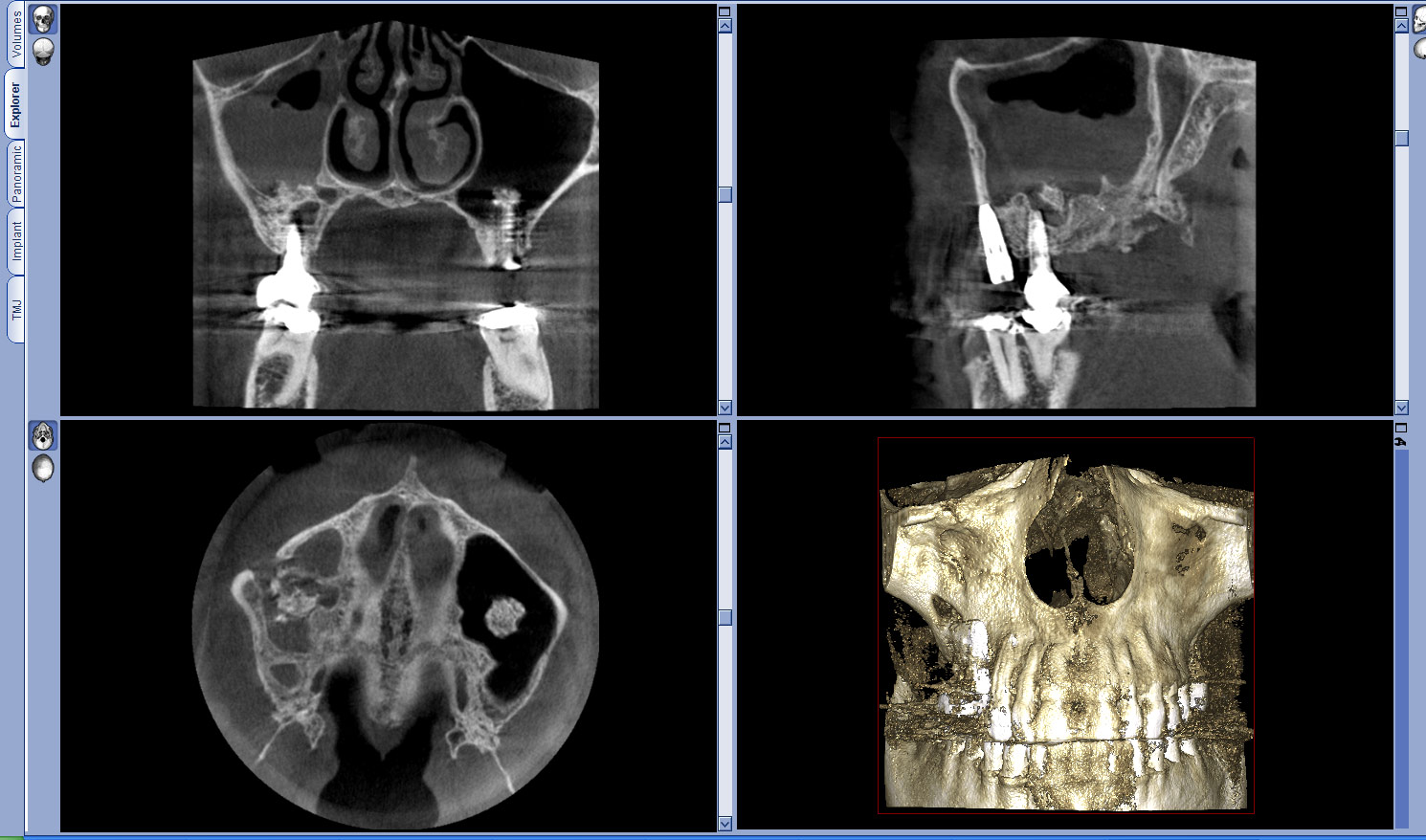
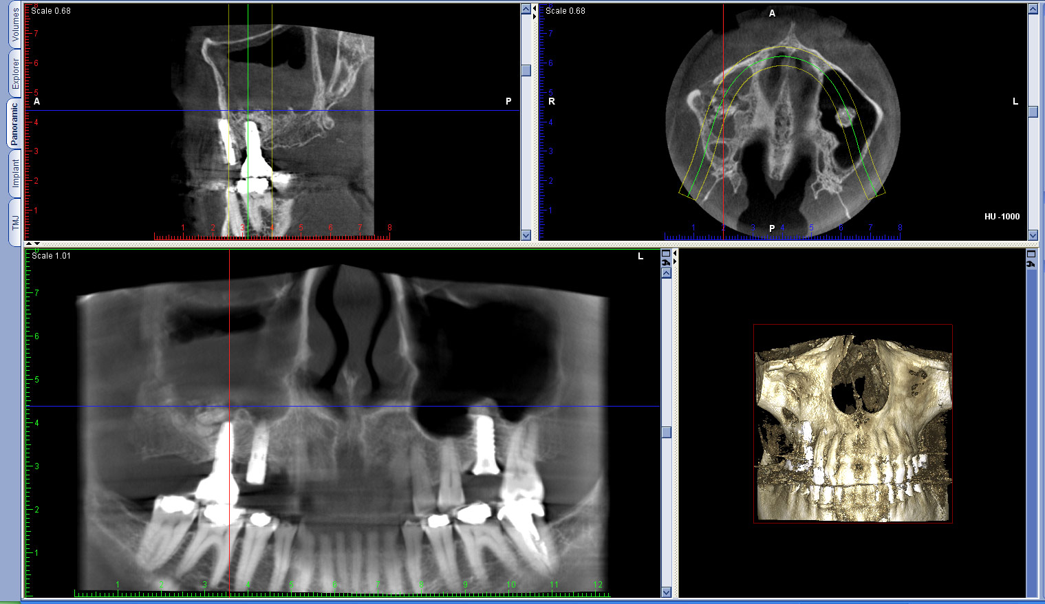
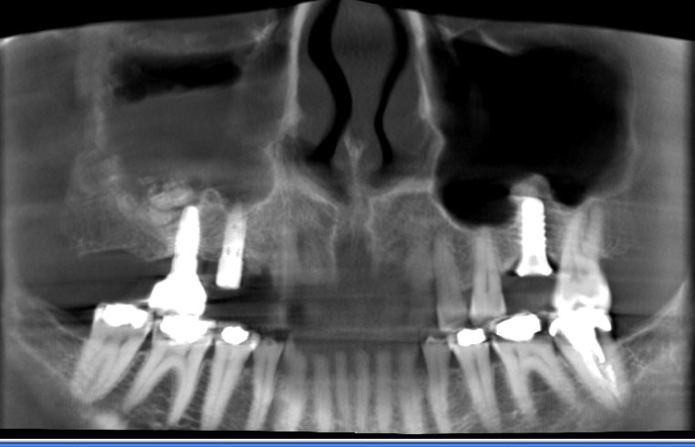
Forum for Clinical and Surgical Oral Pathology Case BBOPF 10-1 Dr. Brian Shumway (University of Louisville, Kentucky, USA) is requesting your opinion. Please send your comments in the window located below the images. This case will be posted from March 6-13, 2010. A summary of the responses will be posted in BBOP. Clinical HistoryThis is a 52yr old caucasian female with a history of right maxillary sinus augmentation with Bio-Oss graft material 2-3 yrs previously. She now presents with a cloudy sinus on PANX. The surgeon reported loose Bio-Oss material and a thickened sinus membrane. The patient has no reported systemic conditions. From these sections this is what I see: chronic sinusitis, bacteria, and fungal microorganisms (I will suggest they culture the patient as per our microbiologist's recommendations). The polarizable and eosinophilic materials are what have me confused. I think that the polarizable material is calcium oxalate (none of the local labs do an oxalate stain- I could send to Mayo if this is necessary to confirm) as it seems similar to what I have seen in the literature and it would go along with the fungus present. The eosinophilic material doesn't look like Bio-Oss from the limited articles I could find that show its histologic appearance. While there were no eosinophils in the biopsy, I did a trichrome as this is reported to highlight Charcot Leydon crystals. The material stains bright red but I am not seeing the bipyramidal forms that are reported. There seem to be many different forms that weren't very evident on H&E. I wondered if this was some other type of graft material and maybe the history was inaccurate. Does anyone know what 2 types of material I may be dealing with and is there a stain that can be done and is it necessary to confirm? Thanks to anyone who can provide insight. Sincerely, Brian Shumway DDS, MS Images(Larger images will open in a new window or tab.)
Case prepared by Dr. Alfredo Aguirre (BBOP Manager) and Daniel Emmer (Web Administrator, University at Buffalo School of Dental Medicine). |