Bulletin Board of Oral Pathology
Bulletin Board of Oral Pathology
|
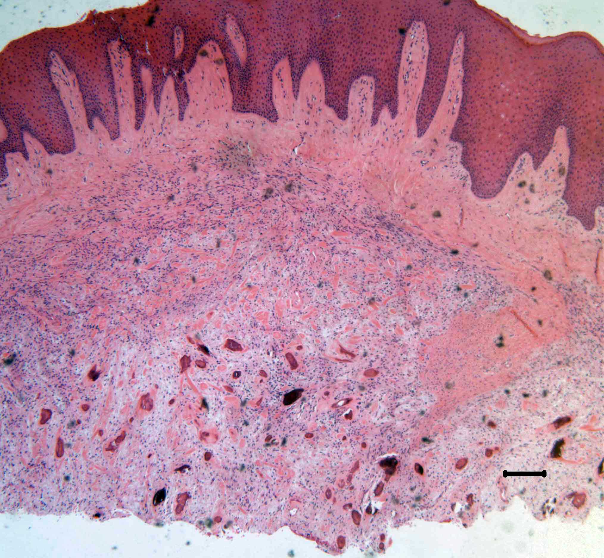
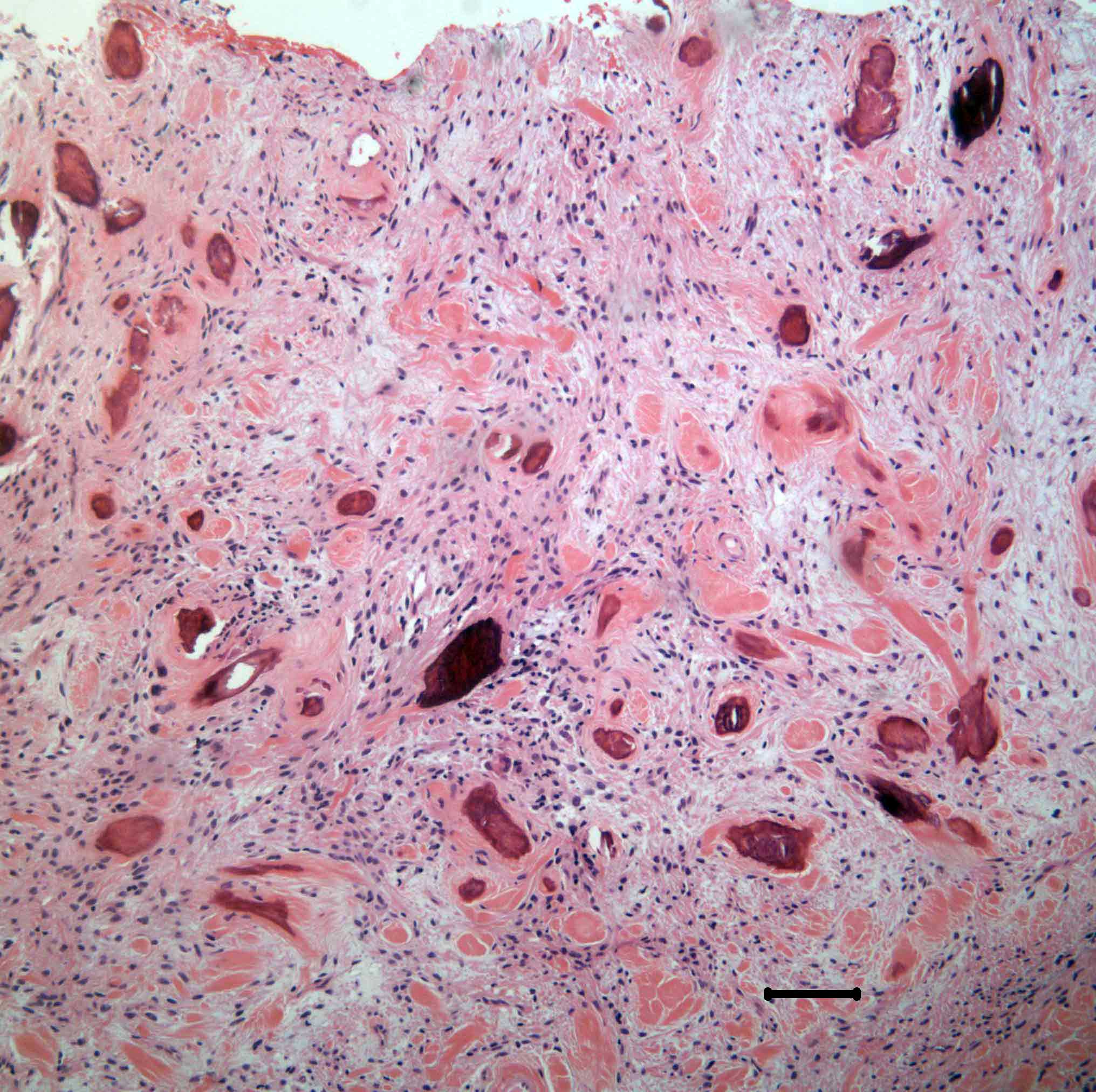
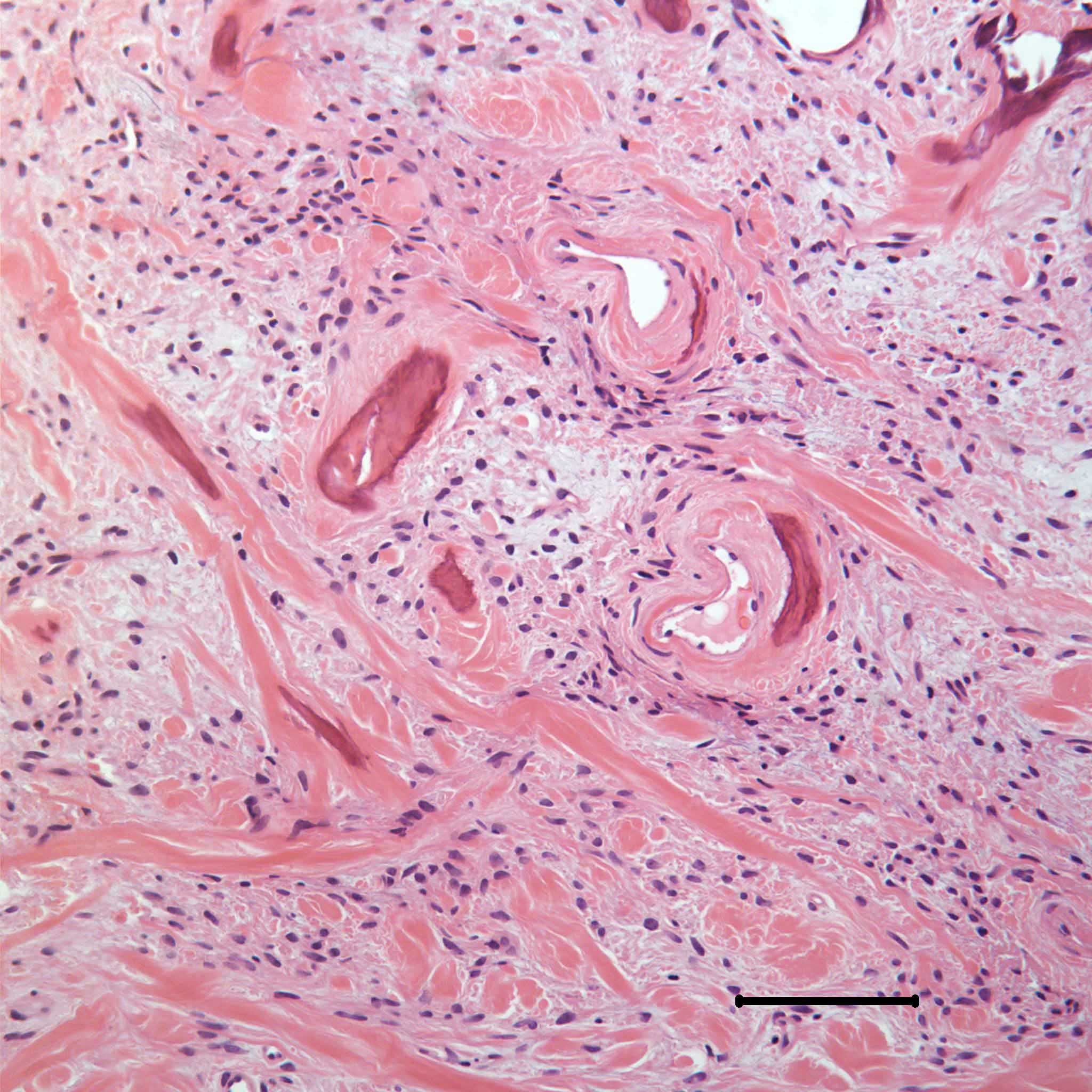
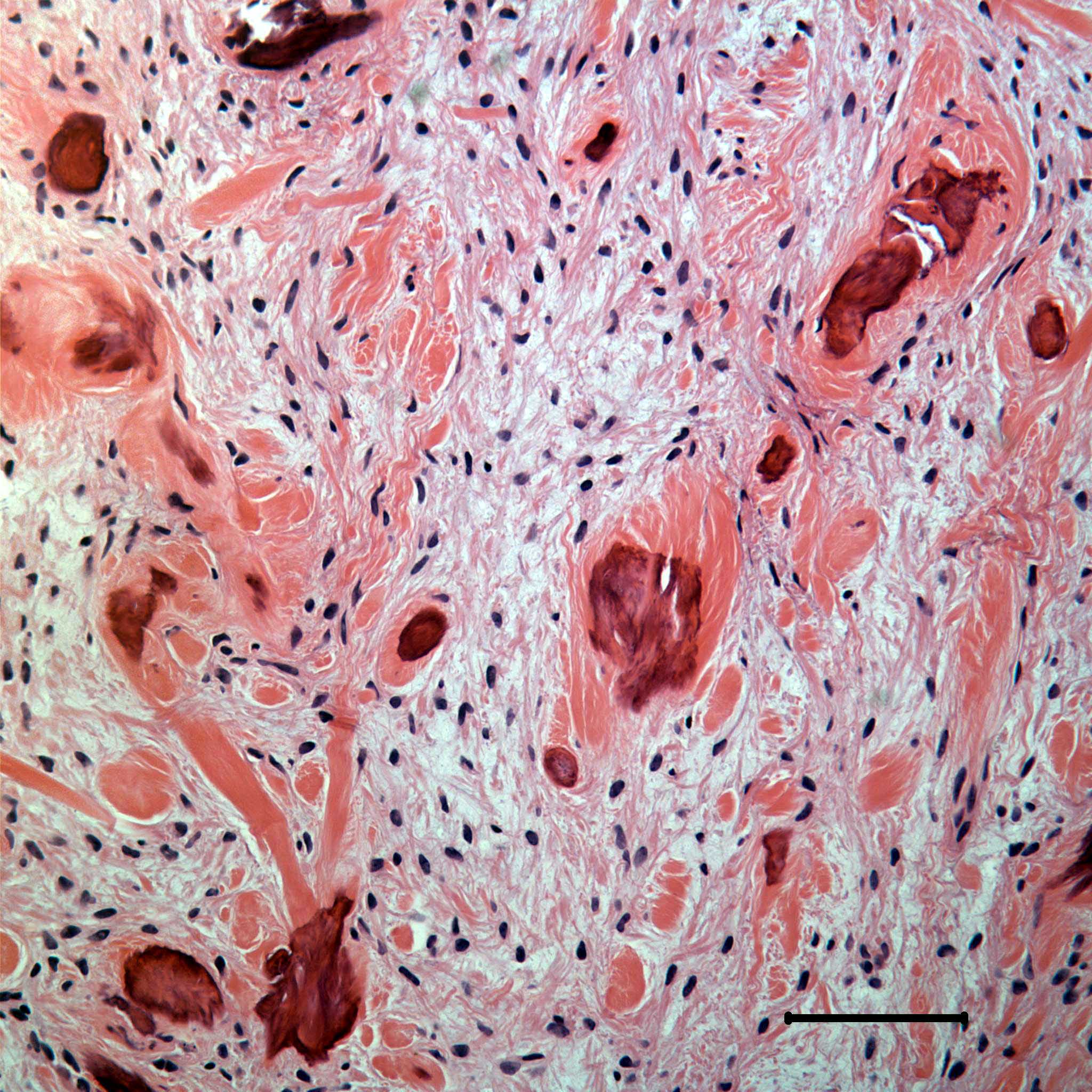
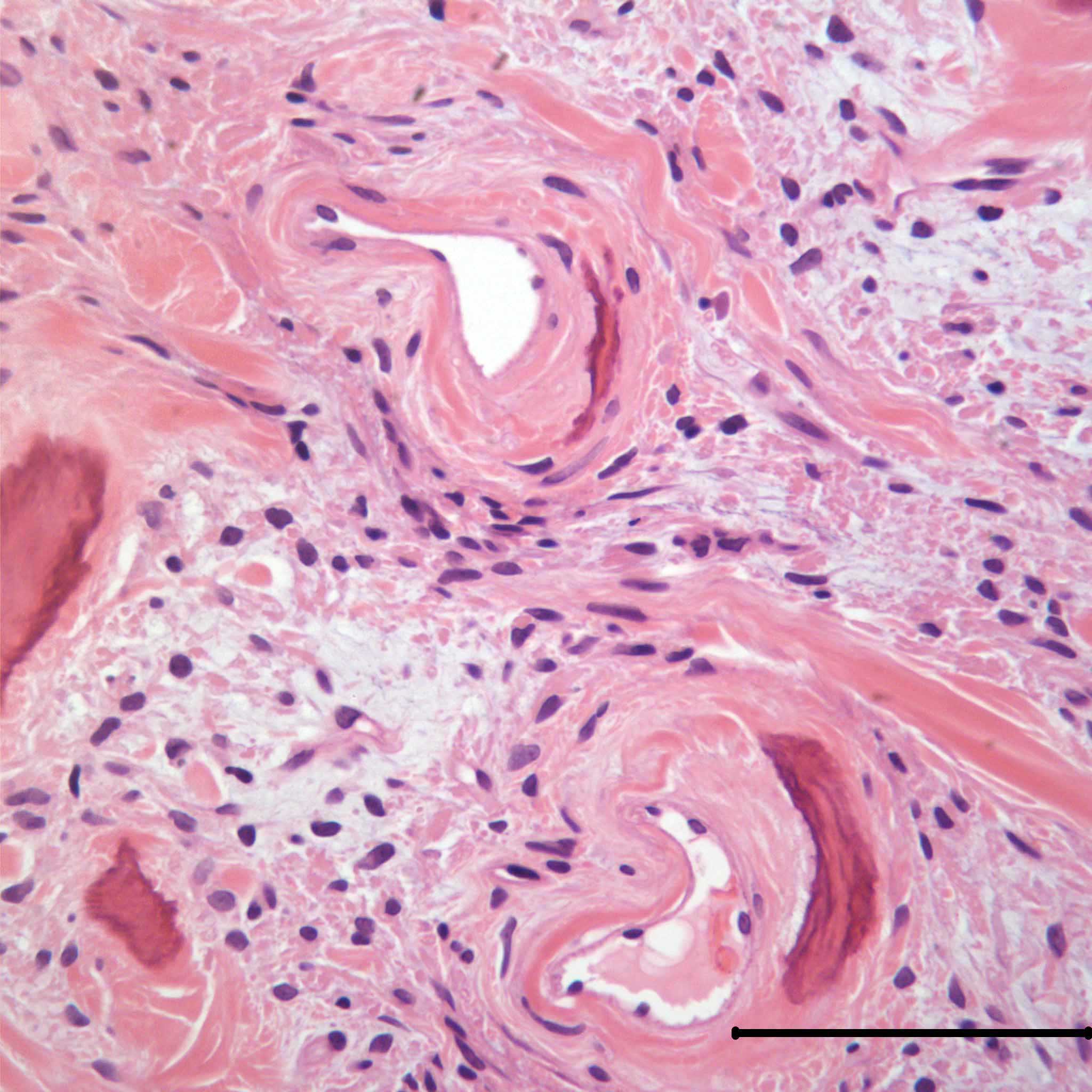
Forum for Clinical and Surgical Oral Pathology Case BBOPF 08-1 Dr. Nadarajah Vigneswaran (The University of Texas Dental Branch at Houston, USA) invites your comments on the microscopic findings of a gingival biopsy. Please send your diagnosis/comments in the window located below the images. This case will be posted from March 3 to March 10, 2008. A summary of the responses will be posted in BBOP. Clinical HistoryA 28 year old female presented with an exophytic lobulated mass (2 x 2 cm) on her mandibular gingiva between teeth # 27 and #28. The lesion involves both facial and lingual gingivae and it has been present for more than one year. Excisional biopsy was performed and the surgeon considered pyogenic granuloma and peripheral ossifying fibroma in his differential diagnosis. Multiple photomicrographs of this biopsy are included for your review and comments. Nadarajah Vigneswaran BDS, DMD, Dr.Med.Dent. Images
Case prepared by Dr. Alfredo Aguirre (BBOP Manager) and Daniel Emmer (Web Administrator, University at Buffalo School of Dental Medicine). |