Bulletin Board of Oral Pathology
Bulletin Board of Oral Pathology
|
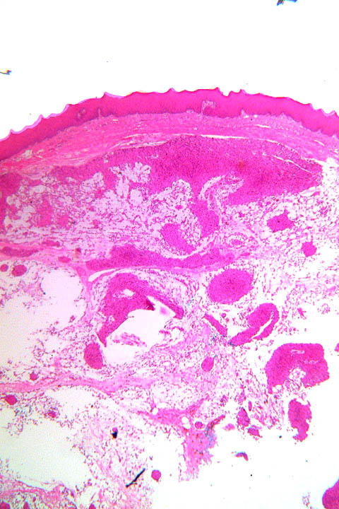
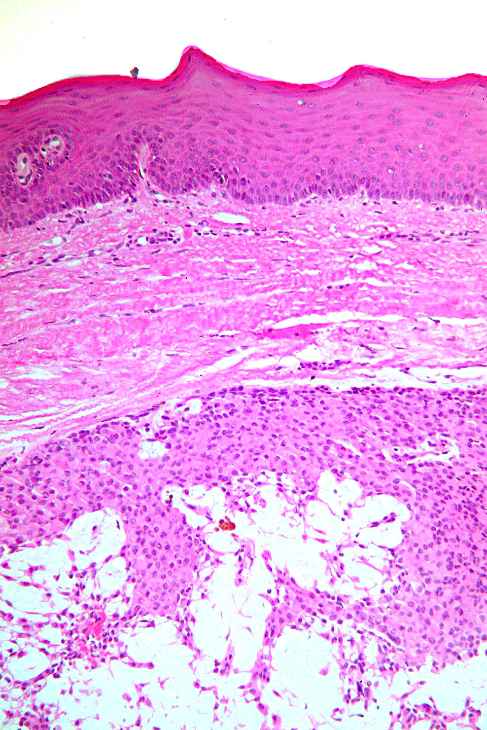
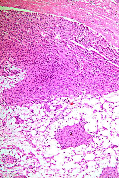
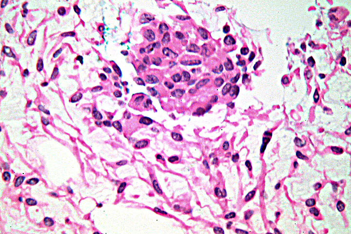
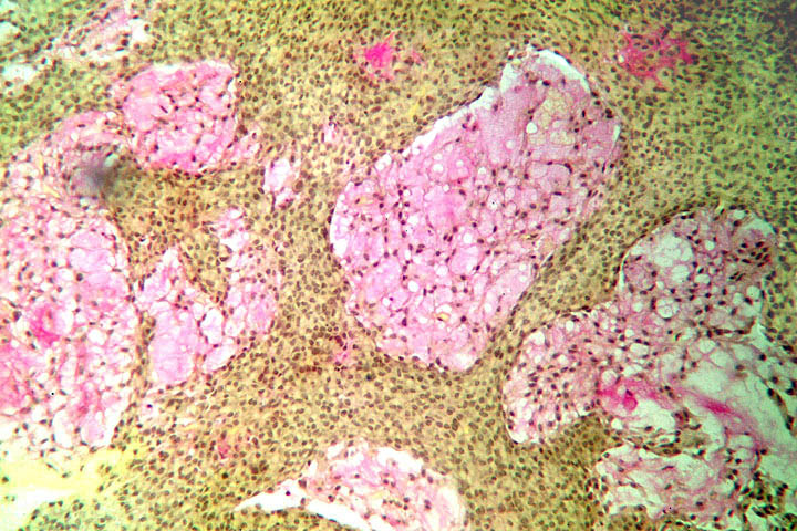
Forum for Clinical and Surgical Oral Pathology Case BBOPF 05-2 The following case was contributed by Dr. Robert Howell (West Virginia University, USA) Dr. Howell invites your comments on the representative microscopic images of one of his current surgical cases. Please send your comments in the window located below the images. This case was posted from March 29 to April 4, 2005. A summary of the responses will be posted in BBOP. Clinical History75 year-old female with a palatal lesion, size was stated as being 1.5 cm. No erosion of bone, but bone was described as being saucerized by pressure. The lesion is of unstated duration. I signed it out as pleomorphic adenoma. However, I have not seen one exactly like this before. The surgical specimen was badly fragmented so I only had one border to exam (under the surface epithelium). I would greatly appreciate your comments. Images
Case prepared by Dr. Alfredo Aguirre (BBOP Manager) and Daniel Emmer (Web Administrator, University at Buffalo School of Dental Medicine). |