Bulletin Board of Oral Pathology
Bulletin Board of Oral Pathology
|
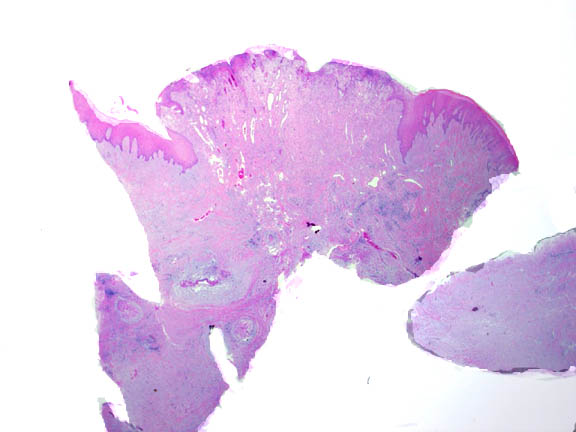
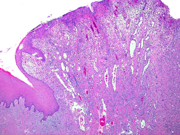
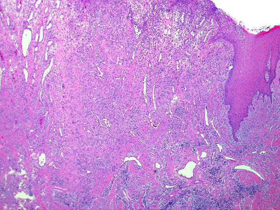
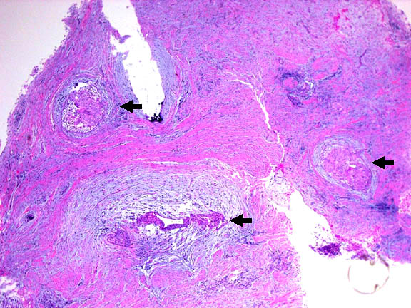
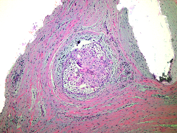
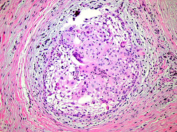
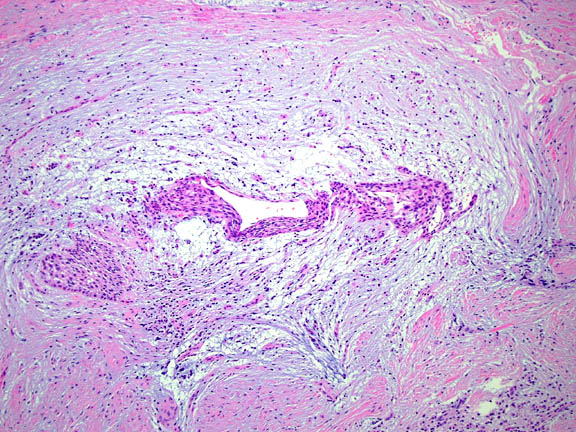
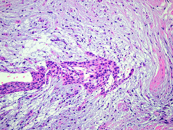
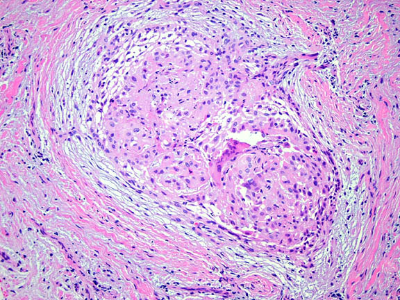
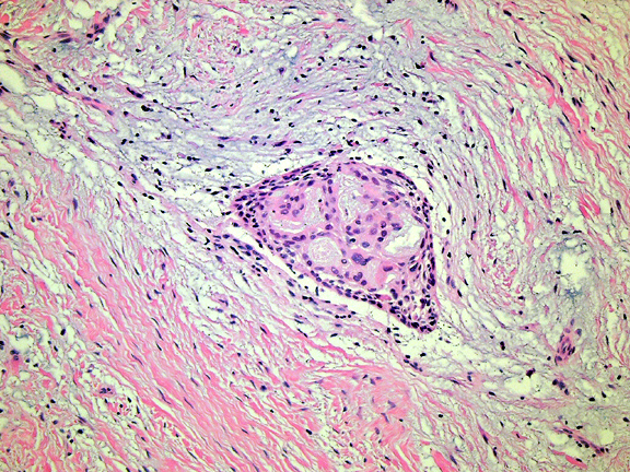
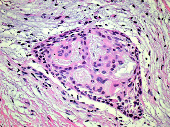
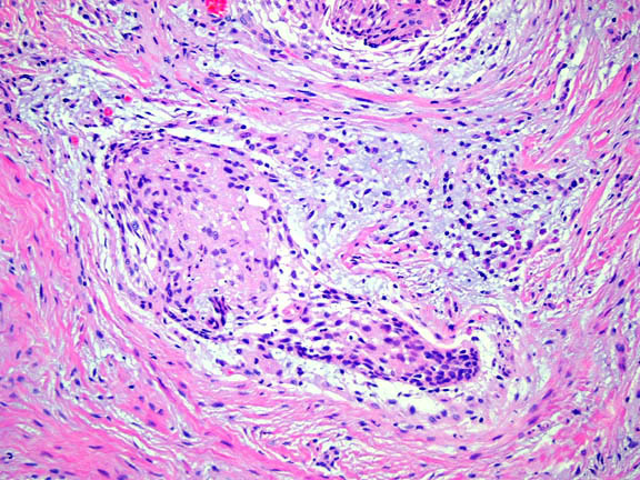
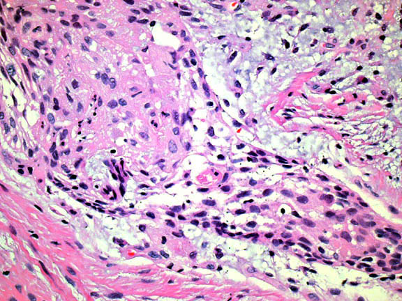
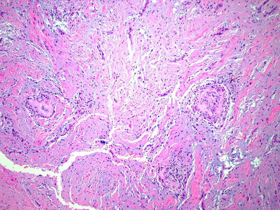
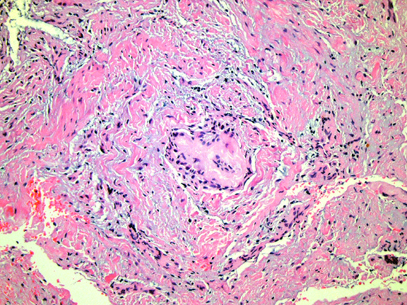
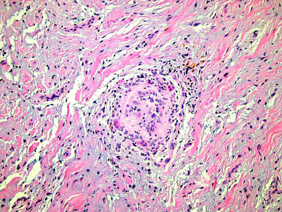
Forum for Clinical and Surgical Oral Pathology Case BBOPF 04-2 Dr. Harvey Kessler invites your comments on the following case. Please send your comments in the window located below the H&E images. This case was posted from February 26 to March 11, 2004. A summary of the responses will be available in BBOP. Clinical HistoryA 42 yo white female had the mandibular right second premolar extracted in December 2003 and has had pain in the site since then. Slightly more than 7 weeks later, a granular exophytic mass was noted in the extraction site. The area continued to be painful. The tissue was removed from the extraction site with the clinical provisional diagnosis of "giant cell granuloma or ossifying fibroma." The superficial portion of the lesion, as can be seen in the photomicrographs, showed pyogenic granuloma and POF areas adjacent to one another. What was different and interesting about the case was in the deeper tissues, removed from the PG/POF areas, where multiple islands of apparent epithelium were seen. The epithelial islands were mildly proliferative and showed some inflammatory exocytosis. In a couple of the islands what appeared to be giant cells were present. In an adjacent fragment of the specimen, foci of "giant cell hyaline angiopathy (GCHA)" were observed, perhaps not unexpected in a prior extraction site. Photomicrographs document the number of islands and the various appearances. GCHA like foci are included for comparison. We interpreted this as proliferation of residual odontogenic epithelial cell rests secondary to the inflammation in the extraction site, but admit that the pattern of proliferation, the cytomorphology, and giant cell presence is unusual in our experience. We did not believe this epithelium was an active component of the lesion although odontogenic fibroma and other odontogenic neoplasms were thoughts too. We are interested to know if others have encountered this same pattern and any opinions/experience as to the significance of these findings are welcomed. Images
|
Case prepared by Dr. Alfredo Aguirre (BBOP Manager) and Daniel Emmer (Web Administrator, University at Buffalo School of Dental Medicine).