Bulletin Board of Oral Pathology
Bulletin Board of Oral Pathology
|
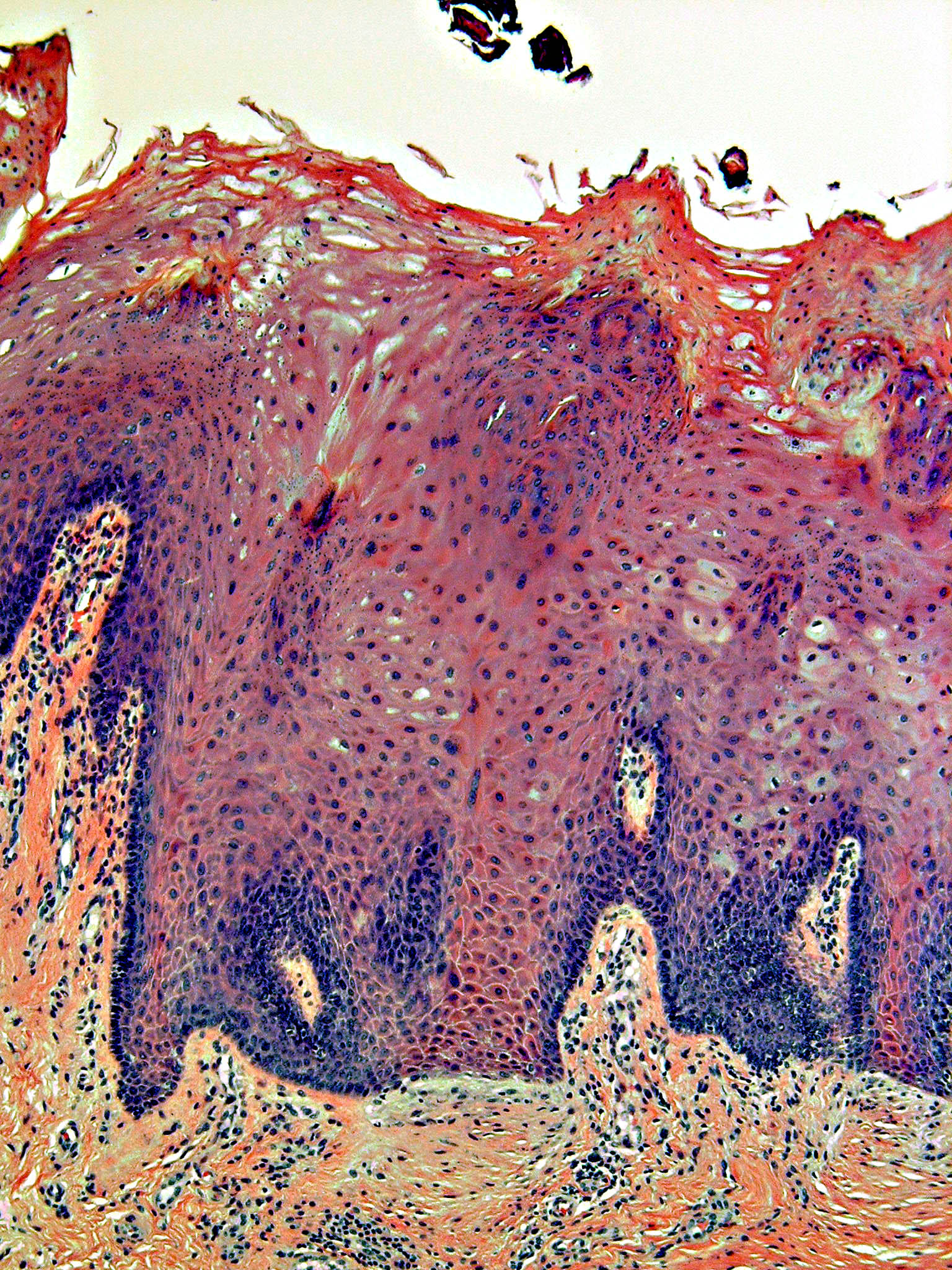
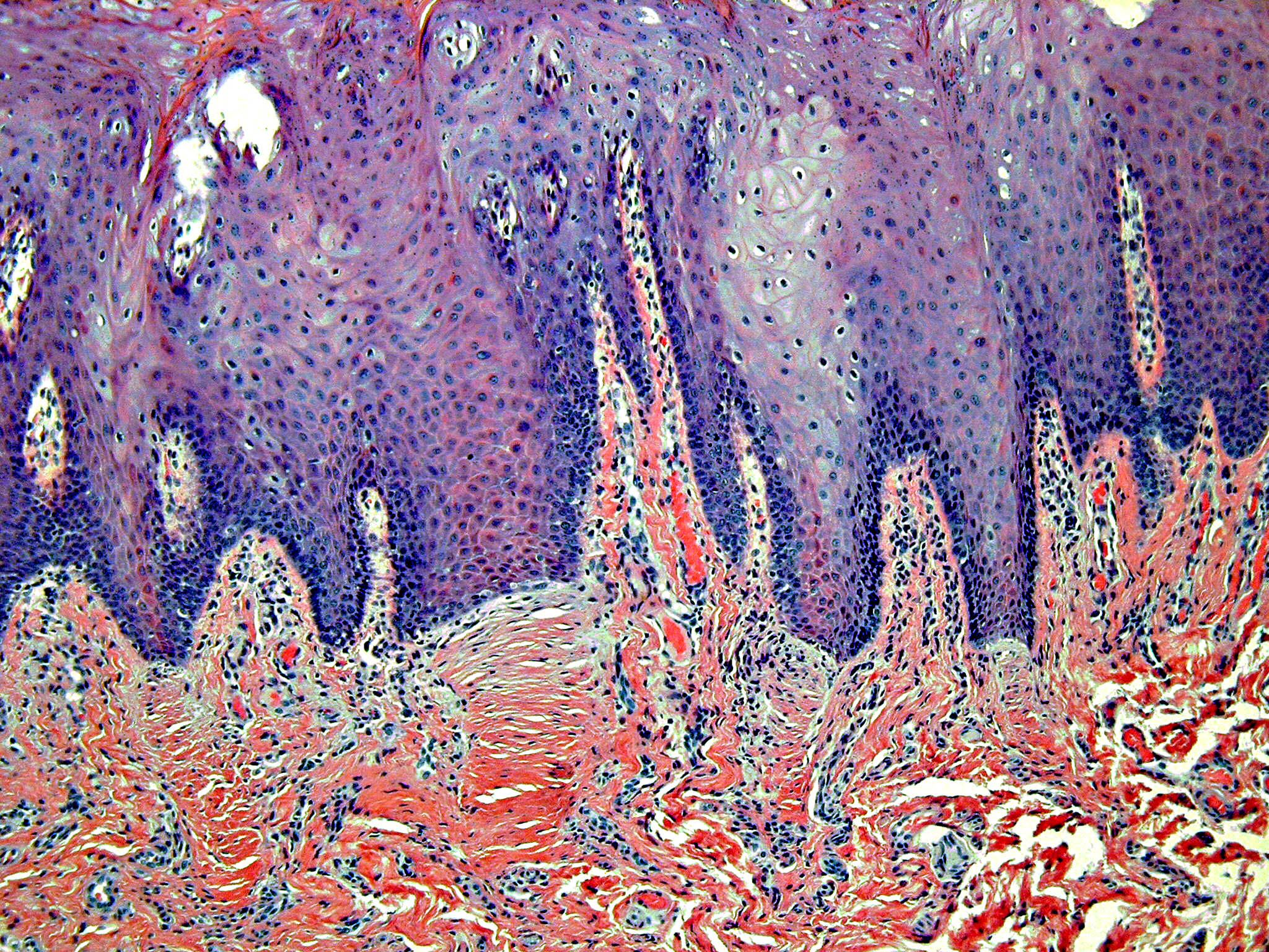
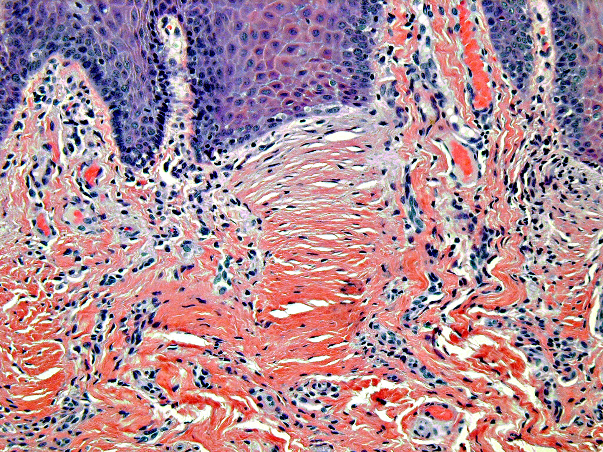
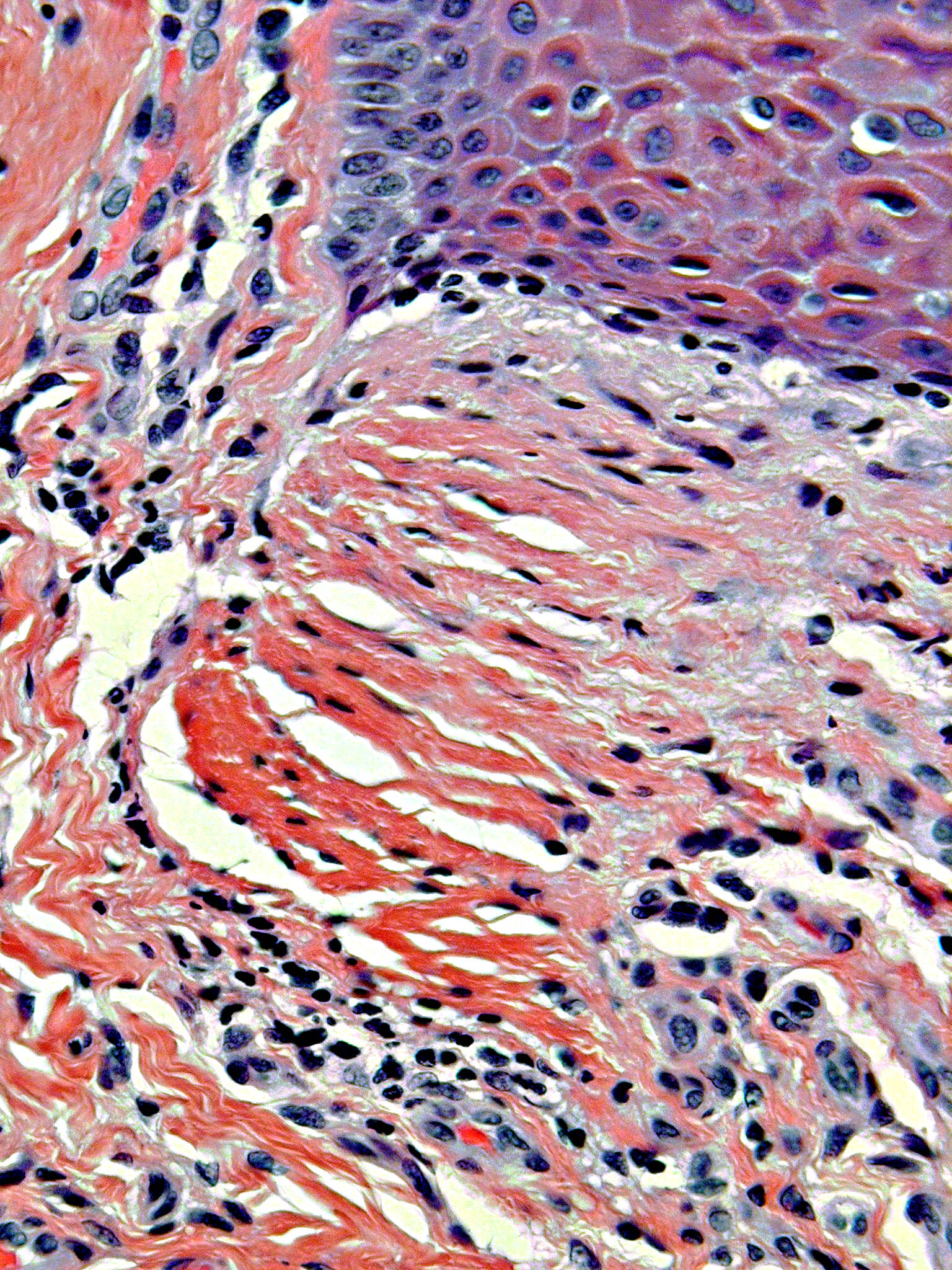
Forum for Clinical and Surgical Oral Pathology Case BBOPF 03-9 The following case was contributed by Dr. John Kalmar (Oral and Maxillofacial Pathology, College of Dentistry, The Ohio State University, Columbus, Ohio, USA). Dr. Kalmar is requesting your comments for the ensuing case, in particular, the collagen fibers arrangement seen underneath a broad based rete peg depicted in the ensuing images (is there a dermatological lesion where this peculiar collagen pattern is observed ?) . Please send your comments in the window located below the images. This case was posted from December 5 to December 19, 2003. A summary of the responses will be posted in BBOP. Clinical History53 yr old female presented with a several week history of sensitive areas of the dorsal tongue. Clinical examination revealed multiple flat circular white spots (1-2 mm) of the mid-dorsal tongue (never quite reaching the lateral border) that seemed to coalesce in some areas, occasionally making U-shaped figures. A biopsy was interpreted by a general pathologist as "consistent with leukoplakia." Representative microscopic images of this case are shown below. Images
Case prepared by Dr. Alfredo Aguirre and Daniel Emmer (Web Administrator, University at Buffalo School of Dental Medicine). | |||||||||||