Bulletin Board of Oral Pathology
Bulletin Board of Oral Pathology
|
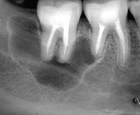
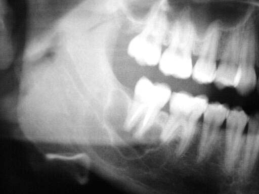
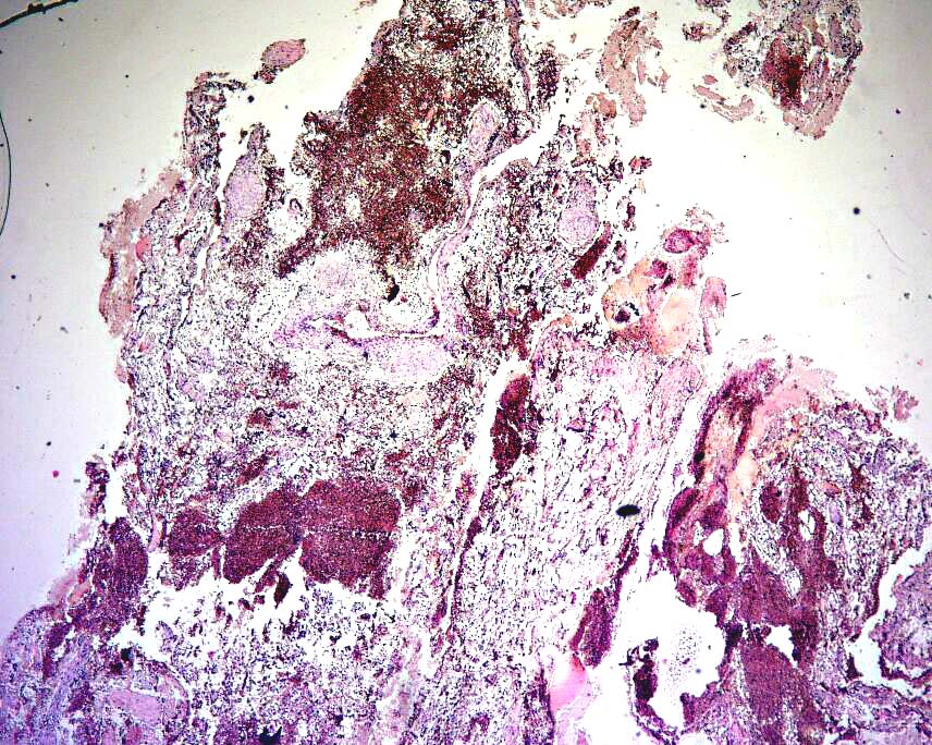
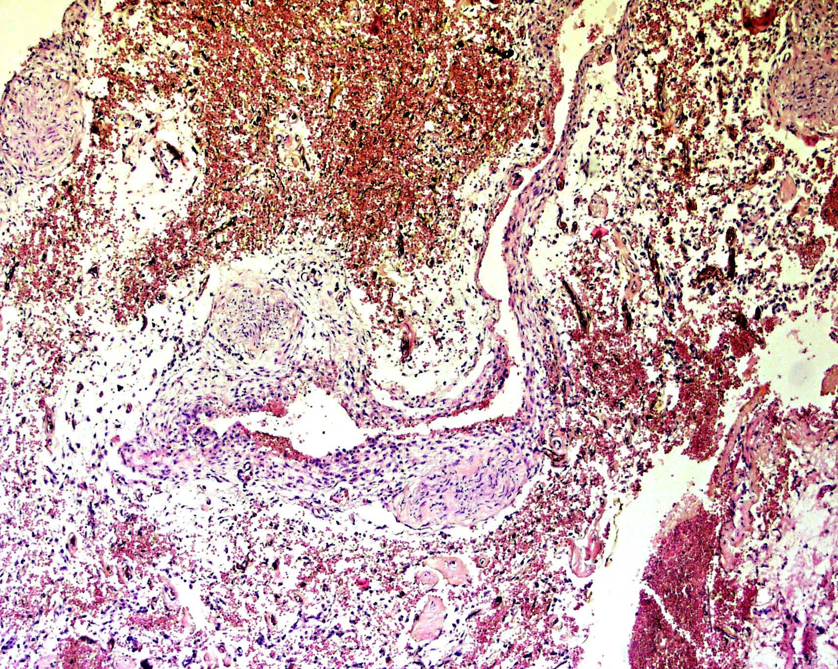
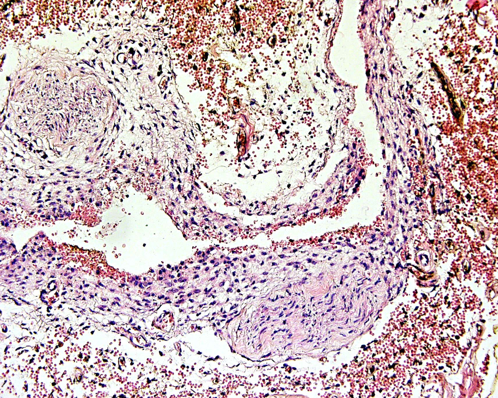
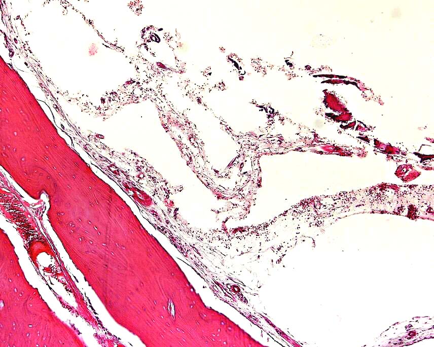
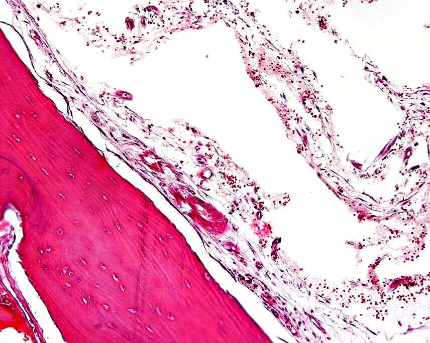
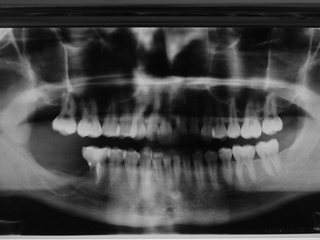
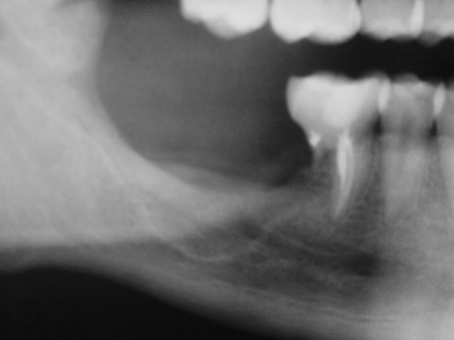
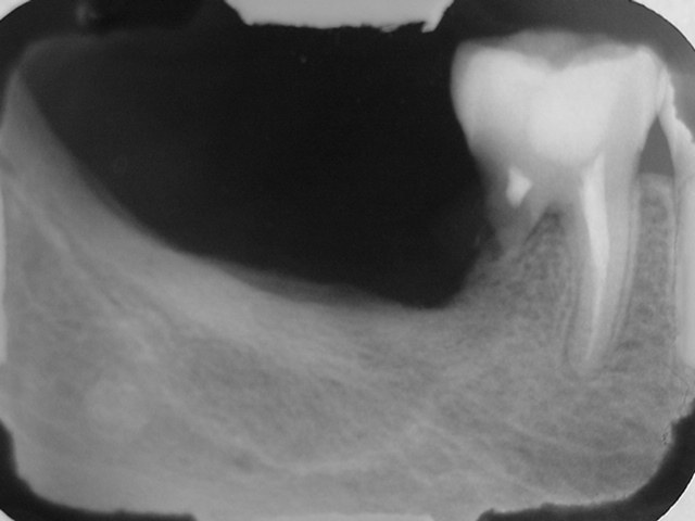
Forum for Clinical and Surgical Oral Pathology Case BBOPF 03-7 The following case was contributed by Dr. Benjamin Martinez (Oral Pathology Department, School of Dentistry, University of Mayor, Santiago de Chile, Chile). Dr. Martinez is requesting your comments for the ensuing case. Please send your diagnosis/suggestions in the window located below the following images. This case will be posted from August 22 to September 5, 2003. A summary of the responses will be posted in BBOP. Clinical HistoryEighteen year-old female presents a corticated radiolucency in the right mandible. A biopsy was performed. The oral surgeon reported that upon entering the lesion an empty cavity was found. Small fragments of soft and hard tissue were obtained after curettage of the lesion. Follow-up of the patient revealed that the radiolucency increased in size. One year after the initial surgical procedure, the area was explored again and presented similar clinical findings as those seen in the first biopsy. The area was curetted and a scarce thin tissue membrane obtained. Three radiographic images of the initial lesion, three H&E stained images representative of the initial surgical specimen and two representative H&E images from the second biopsy are included. Images
Case prepared by Dr. Alfredo Aguirre (BBOP Manager) and Daniel Emmer (Web Administrator, University at Buffalo School of Dental Medicine). | |||||||||||||||||||||||