|
Forum for Clinical and Surgical Oral Pathology
Case BBOPF 03-4
The following case was contributed by Dr. Janett Soriano and Dr. Beatriz Aldape
from the Oral Pathology Department, Faculty of Odontology, National Autonomous
University of Mexico. Drs. Soriano and Aldape are requesting your comments
for the ensuing case. Please send your diagnosis/suggestions in the window
located below the following images. The case was posted from June 10 to
June 21, 2003. A summary of the responses will be posted in BBOP.
Clinical History
62 years-old healthy female without any significant past medical history sought
dental care for replacement of a mandibular removable partial denture. Oral
examination revealed an edentulous maxilla. Teeth # 22-28 were present in the
mandible. In addition, a swelling with a bluish discoloration, well-defined
borders and smooth surface measured about 1.0 cm in its largest diameter (Figure
1). The lesion was asymptomatic and the patient was not aware of it. A panoramic
film was obtained and revealed a bone concavity corresponding to the area of
the clinical lesion. The bone cortex seemed to be intact (Figure 2).
An excisional biopsy was obtained. Five µm paraffin embedded sections
were obtained and stained with H&E (Figures 3-13). In addition, PAS and
alcian blue stains were also obtained (Figures 14 and 15).
Images
Click on images to open a larger version in another browser window.
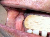
Figure 1
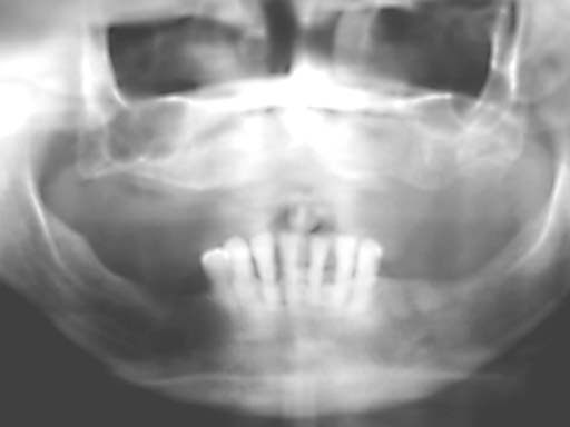
Figure 2
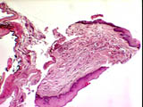
Figure 3 x 10 |
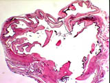
Figure 4 x 10 |
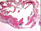
Figure 5 x 10 |
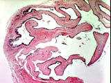
Figure 6 x 100 |
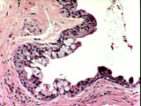
Figure 7 x 100 |
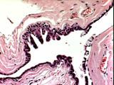
Figure 8 x 100 |
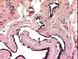
Figure 9 x 100 |
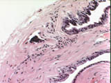
Figure 10 x 100 |
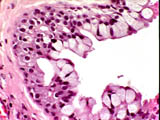
Figure 11 x 200 |
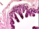
Figure 12 x 200 |
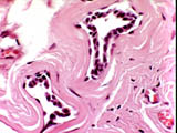
Figure 13 x 200 |
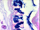
Figure 14 Alcian Blue x 200 |
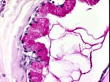
Figure 15 PAS x 200 |
THIS IS AN ACADEMIC EXERCISE ONLY. THE REPLIES ARE NOT TO BE CONSTRUED AS THOSE OF A CONSULTANT FOR LEGAL PURPOSES.
|