Bulletin Board of Oral Pathology
Bulletin Board of Oral Pathology
|
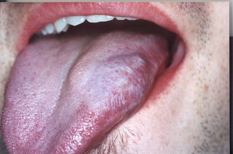
Forum for Clinical and Surgical Oral Pathology Case BBOPF 02-4 The following case was contributed by Dr. Paul Botha (private practice in periodontology, Cape Town, South Africa). This case was posted from December 6 to December 20, 2002. A summary of the responses will be available here. Case History20-year old man reported with a unilateral white lesion on the dorso-lateral aspect of the left side of the tongue which has been present for 3 months. The lesion is not painful but sensitive to spicy foods. He is a very fit man who works out in the gym and had previously been consuming a dietary supplement. He works as a printing apprentice and is exposed to paraffin, alcohol and ink on a daily basis. He does not smoke or drink or use recreational or other drugs. He uses Aquafresh toothpaste with the red stripe. Clinical examination revealed a 4cm x 1,5cm white lesion which could not be rubbed off. The fungiform papillae are still readily identifiable in the lesion with the rest of the lesion having cells which seem 'bloated'. On the clinical photo the lesion seems raised but clinically it is not. Biopsy report was sent from the maxillo-facial surgeon who consulted him initially and the following report was sent from a board-certified oral pathologist: Acanthotic squamous epithelium covered with parakeratin. Large, pale surface cells indicate possible trauma. There are lengthened rete ridges and their are signs of basal cell degeneration as well as an interface mucocitis. The basal membrane is irregularly thickened. The lamina propia is replaced by granulation tissue. A band-like infiltrate composed of lymphocytes and macrophages is present under the epithelium. No candida could be seen with special staining. The microscopic features are suggestive of trauma. Lichen planus or lichenoid reaction can also be included in a differential diagnosis. The following tests were also negative:
Initial treatment consisted of withdrawing his toothpaste for 2 weeks. This did not cause any change in the lesion. Topical steroid treatment applied twice per day for 2 weeks did not reveal any change. ImagesClick on image to open a larger version in another browser window.
Case prepared by Dr. Alfredo Aguirre (BBOP Manager) and Daniel Emmer (Web Administrator, University at Buffalo School of Dental Medicine). |