Bulletin Board of Oral Pathology
Bulletin Board of Oral Pathology
|
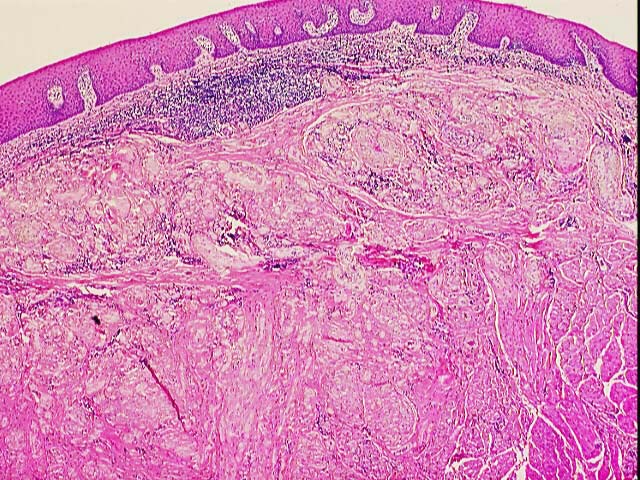
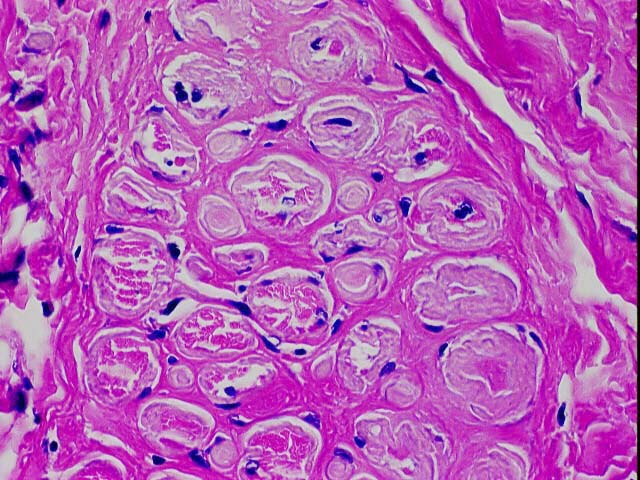
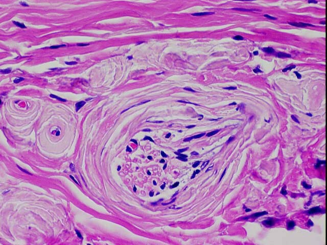
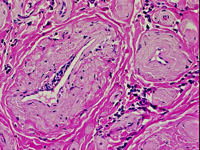
Forum for Clinical and Surgical Oral Pathology Case BBOPF 02-1 The following case was contributed by Dr. Douglas Damm. This case was posted from October 22 to October 31, 2002. A summary of the responses will be available here soon. A 25 year old Caucasian female presented with a firm submucosal nodule of the ventral surface of the tongue. The lesion was asymptomatic and of unknown duration. An excisional biopsy was performed. In a central wedge-shaped zone, the specimen reveals deposition of an eosinophilic and amorphous material that closely resembles amyloidosis. The material extends from the superficial lamina propria to deep within the underlying muscle. It concentrically surrounds all of the blood vessels and nerves in the affected zone. It also demonstrates a most unique pattern in the muscle. The cross section of each muscle fiber resembles a bulls eye. The center circle consists of vital muscle surrounded by a definite ring of the amorphous material; both of these layers are surrounded by the sarcolemma. The area is not coagulative necrosis; the nuclear staining remains intense. The patient does not use smokeless tobacco. The clinical presentation and histopathologic features do not support amyloidaceous deposits in ligneous conjunctivitis. The Congo red stains were negative (not amyloid). PAS stains were negative (not lipoid proteinosis, colloid degeneration, amyloid-like deposits of immunotactoid glomerulopathy). Does this represent some unusual pattern of local tissue degeneration similar to the colloid degeneration that occurs in sun damaged skin? HELP. Anybody know of an amyloid-like deposit that is negative on Congo Red and PAS? ImagesClick on images to open a larger version (2X) in another browser window.
Case prepared by Dr. Alfredo Aguirre (BBOP Manager) and Daniel Emmer (Web Administrator, University at Buffalo School of Dental Medicine). |