Bulletin Board of Oral Pathology
Bulletin Board of Oral Pathology
|
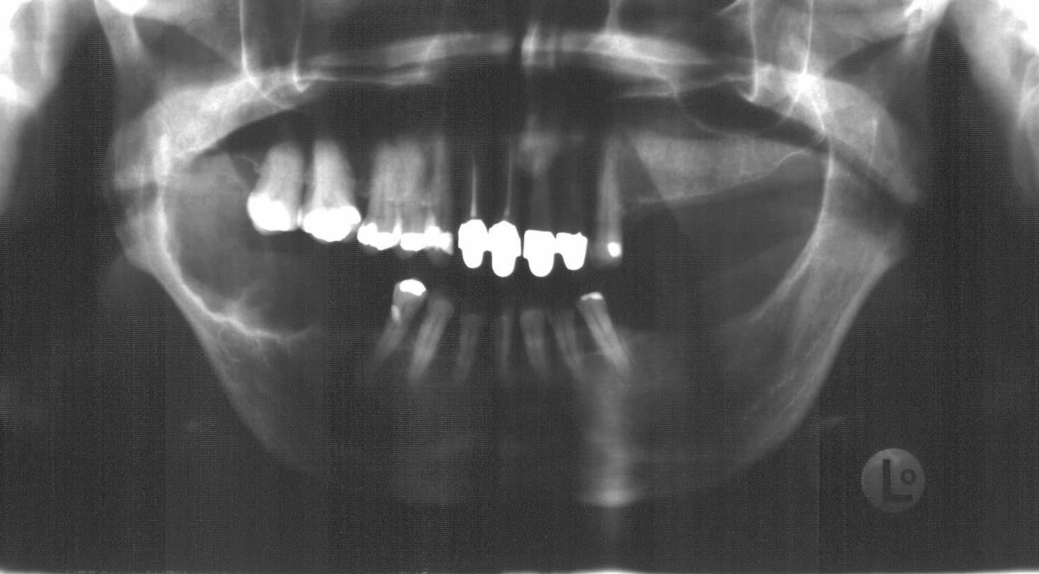
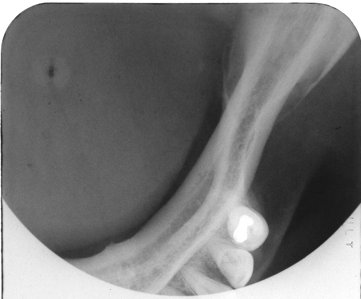
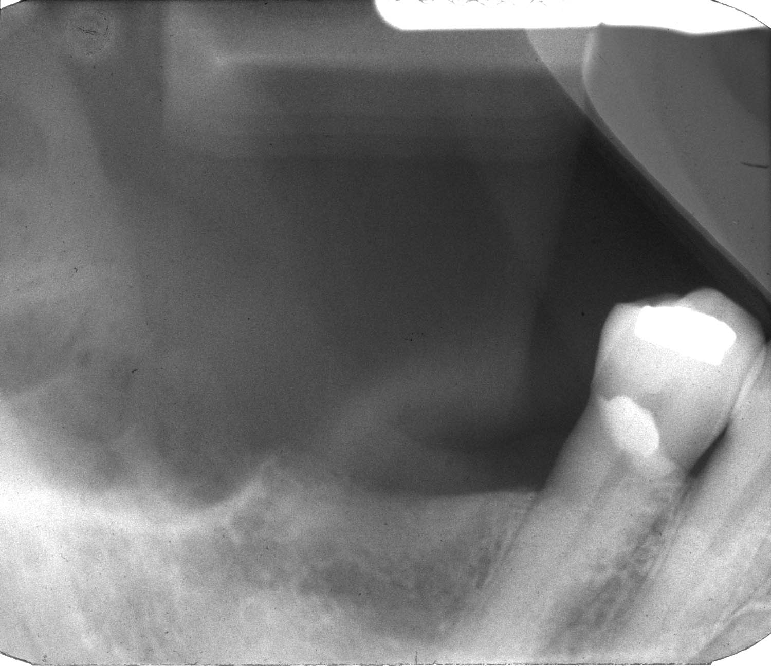
Forum for Clinical and Surgical Oral Pathology Case BBOPF 01-1 The following case was contributed by Dr. John Lovas. This case was posted from July 10 to August 10, 2001. A summary of the responses is available here MAL is a 62 year-old female. CC: "Sore lump" of the right posterior mandible, first noticed by the patient around 2 months ago, with apparently no change since. HPC: The teeth in the area were extracted by her GP dentist over 9 yrs ago - she does not recall any unusual treatment or problems. PMH: Systemic sclerosis diagnosed 11 yrs ago (has Raynaud's, esophagus dilated 3x); hospitalized 1 yr ago for Stevens-Johnson syndrome (erythema multiforme major) - "I'm allergic to many things"; 2 transient ischaemic attacks 7 months ago; has had 8 kidney stones; takes felopidine, ramipril, levothyroxine, hydrochlorothiazide; quit smoking (socially x 5 yrs) 22 yrs ago. PE: Examination revealed no regional lymphadenopathy; right posterior mandible large oval buccal (~3 cm) / occlusal cortical expansion from the edentulous second premolar area, up to & including the mandibular ramus. The overlying mucosa is intact, but slightly bluish focally, suggestive of underlying fluid content. There's no current clinical evidence of infection / inflammation. RADIOGRAPHS: Scans of panoramic ("MALpan"), occlusal ("MALocclu") and periapical ("MALpa") radiographs are included. Click on images to open a larger version (3X) in another browser window.
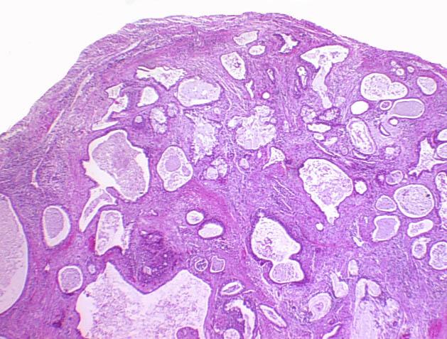
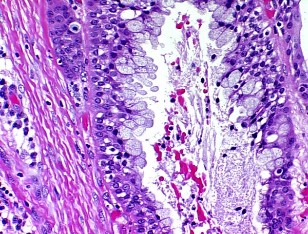
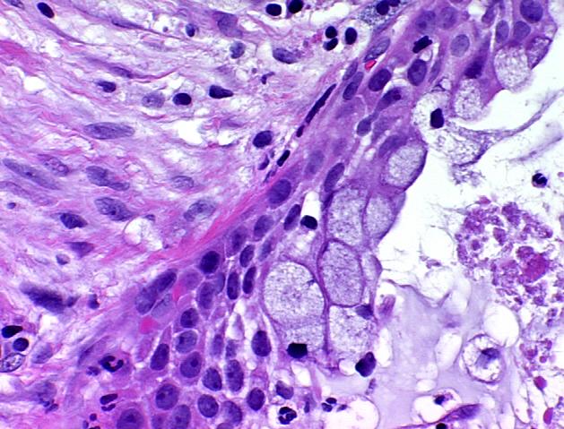
Malpan DISP: She has been referred to an OMF Surgeon, CT scans have been ordered, and she is booked for an incisional biopsy. Biopsy Results (click on images to open a larger version (2X) in another browser window):
Case prepared by Dr. Alfredo Aguirre, Dr. Alan Drinnan (BBOP Managers) and Daniel Emmer (Web Administrator, University at Buffalo School of Dental Medicine). |