Bulletin Board of Oral Pathology
Bulletin Board of Oral Pathology
|
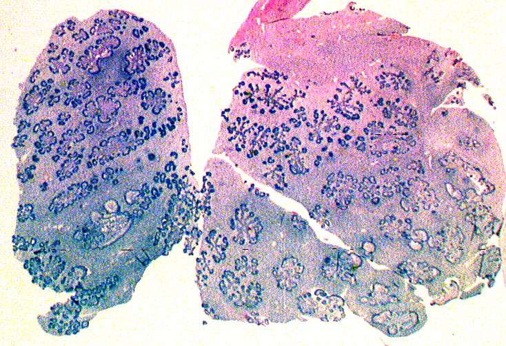
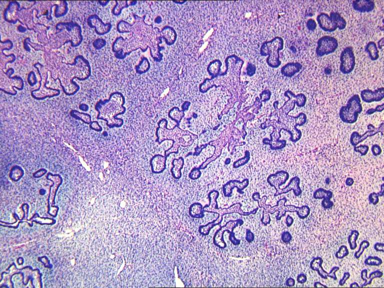
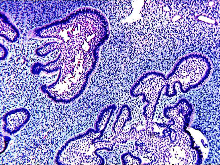
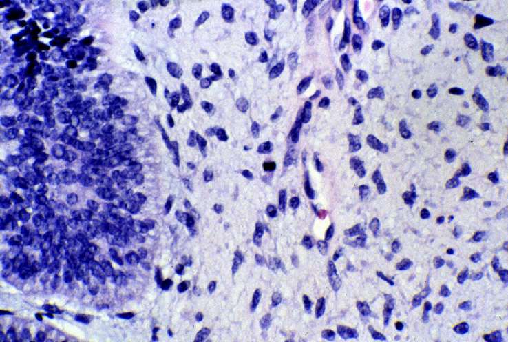
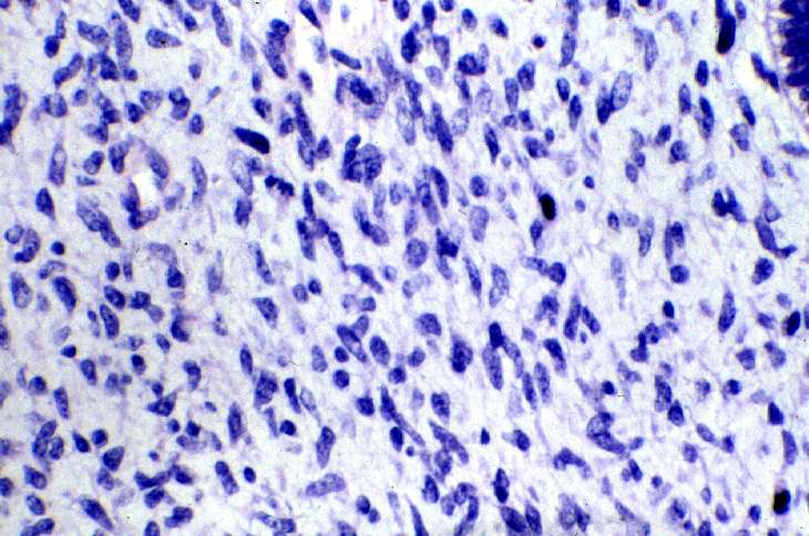
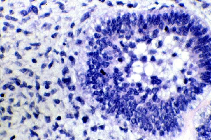
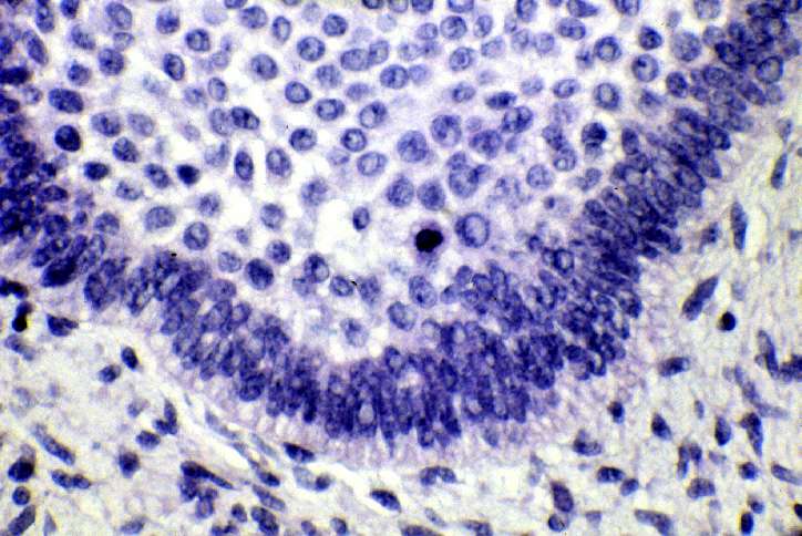
Forum for Clinical and Surgical Oral Pathology The following case originated from upstate New York. The case was be posted from July 27 to August 16, 1998. A summary of the responses is available here. Clinical HistoryA 10 month-old girl presented with an expansile 1.5 cm firm mass of the left anterior maxilla. Radiographically, the tumor was lucent, primarily unilocular and exhibited expansile borders although a small erosion through the palate was indicated. Developing tooth buds noted on the contralateral side seemed to be caught up in this process. The following are 8 representative microscopic fields (stained with H&e) of the
tumoral mass. Clicking any small image will present a much larger version of the image in a separate browser window. After viewing an image in this separate window, you may close, hide, or minimize that window and return to this page. 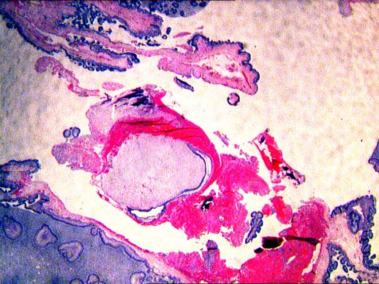 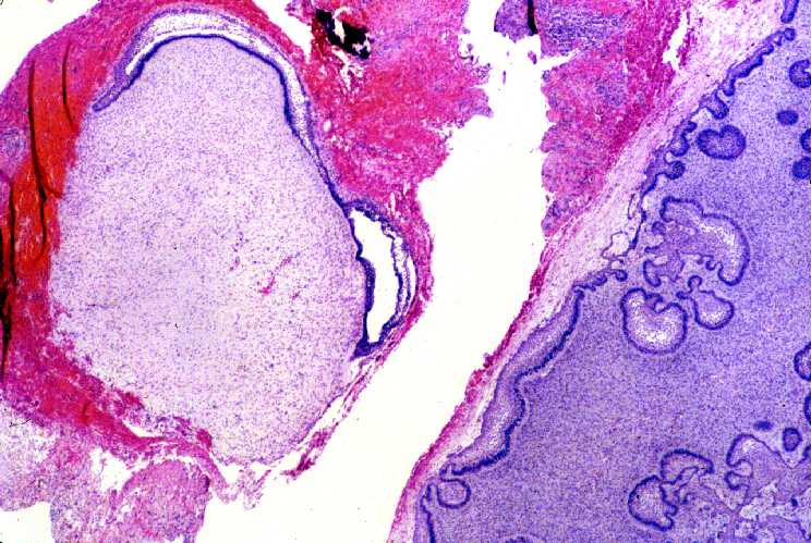 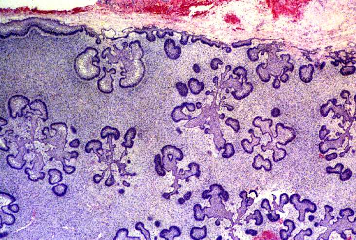
[click for larger images]
|
|
Case prepared by Dr. Alfredo Aguirre, Dr. Alan Drinnan (BBOP Managers) and Daniel Emmer (Web Administrator, University at Buffalo School of Dental Medicine).
|